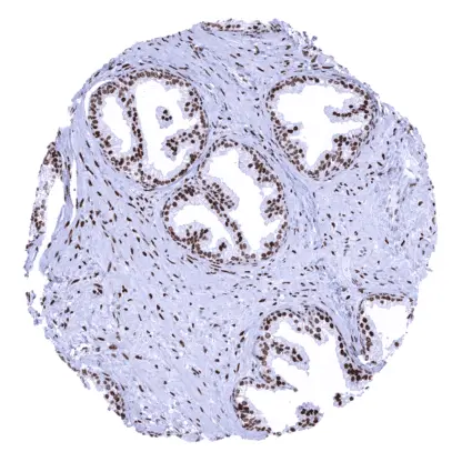
Recombinant Rabbit monoclonal / IgG 1:100 – 1:200 Research Use Only Intracellular Human HMV325 anterior gradient 2, protein disulphide isomerase family member , AG-2 , AG2 , GOB-4 , HAG-2 , HEL-S-116 , HPC8 , PDIA17 , XAG-2 Colon: A strongpredominantly cytoplasmic AGR2staining should be seen in all epithelial cells. Colon:AGR2staining should be absent in all non-epithelial cell types. AGR2 is a catalysator of proper protein folding. Anterior gradient protein 2 homolog (AGR-2), also known as secreted cement gland protein XAG-2 homolog, is a 20 kDa protein disulfide isomerase (PDI) which is coded by the AGR2 gene on chromosome 7p21. AGR2 is a resident endoplasmic reticulum protein which plays an important role in oxidative protein folding in the endoplasmic reticulum. It is expressed in many different tissues/cell types and exerts functions that are relevant for embryonal development, mucus maturation, tissue regeneration, and wound healing. AGR2 is critical for maintaining epithelial barrier function in the intestine. AGR2 knockout mice show a loss of intestinal mucus and develop ileitis and colitis. AGR2 upregulation (and downregulation) has been associated with tumor initiation, progression, and metastasis in several cancer types. Images describing the AGR2 staining pattern in normal tissues obtained by the antibody HMV325 are shown in our “ Normal Tissue Gallery ”. Brain Cerebrum Negative. Cerebellum Negative. Endocrine Tissues Thyroid Negative. Parathyroid Negative. Adrenal gland Negative. Pituitary gland Predominantly cytoplasmic AGR2 staining of variable intensity in variable fractions of epithelial cells of the adenohypophysis. Respiratory system Respiratory epithelium Strong, predominantly cytoplasmic AGR2 staining of all epithelial cells. Lung Strong, predominantly cytoplasmic AGR2 staining of a large subset of pneumocytes. Gastrointestinal Tract Salivary glands Moderate to strong, predominantly cytoplasmic AGR2 staining of subsets of glandular cells (especially mucinous). Weak to moderate AGR2 staining of a subset of excretory duct cells. Esophagus Negative. Stomach Strong, predominantly cytoplasmic AGR2 staining of all epithelial cells except parietal cells. Duodenum Strong, predominantly cytoplasmic AG... AGR2 is most expressed in gastrointestinal, breast and lung cancer, but can also be seen in various other tumor entities. The TCGA findings on AGR2 RNA expression in different tumor categories have been summarized in the Human Protein Atlas. Non-invasive urothelial carcinoma (low grade,
pTaG2) with strong AGR2 positivity of all tumor
cells. AGR2 negative muscle-invasive urothelial
carcinoma. Invasive lobular breast cancer with strong AGR2
positivity of tumor cells. Cancer tissue gallery No data available at the moment IHC users have different preferences on how the stains should look like. Some prefer high staining intensity of the target stain and even accept some background. Others favor absolute specificity and lighter target stains. Factors that invariably lead to more intense staining include higher concentration of the antibody and visualization tools, longer incubation time, higher temperature during incubation, higher temperature and longer duration of the heat induced epitope retrieval (slide pretreatment). The impact of the pH during slide pretreatment has variable effects and depends on the antibody and the target protein. All images and data shown here and in our image galleries are obtained by the manual protocol described below. Other protocols resulting in equivalent staining are described as well. Manual protocol Freshly cut sections should be used (less than 10 days between cutting and staining). Heat-induced antigen retrieval for 5 minutes in an autoclave at 121°C in pH 7,8 Target ... The diagnostic, prognostic, and predictive role of AGR2 expression in tumors and in preneoplastic disease needs to be clarified. There are two ways how the specificity of antibodies can be documented for immunohistochemistry on formalin fixed tissues. These are: 1. Comparison with a second independent method for target expression measurement across a large number of different tissue types (orthogonal strategy), and 2. Comparison with one or several independent antibodies for the same target and showing that all positive staining results are also seen with other antibodies for the same target (independent antibody strategy). Orthogonal validation: For the antibody HMV325 specificity for detection of AGR2 is suggested by the strong concordance of the immunostaining data with data from three independent RNA screening studies, including the Human Protein Atlas (HPA) RNA-seq tissue dataset, the FANTOM5 project, and the Genotype-Tissue Expression (GTEx) project, which are all summarized in the Human Protein Atlas (Tissue expression AGR2) . AGR2 positivity by HMV325 is detectable in all tissues with documented AGR2 RNA...