p16 (MSVA-016R)
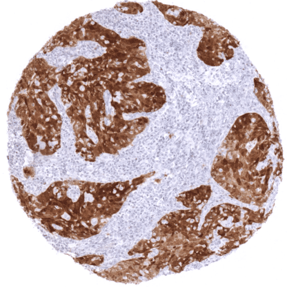
Recombinant Rabbit monoclonal / IgG 1:100 – 1:200 Research Use Only Nuclear and Cytoplasmic Human MSVA-016R CDK4 inhibitor p16 INK4; CDK4I; CDKN2A; Cell cycle negative regulator beta; CMM2; Cyclin dependent kinase 4 inhibitor A; Melanoma p16 inhibits CDK4; MLM; MTS1; Multiple tumor suppressor 1; p14; p16; p19; P19ARF; TP16 Pancreas: at least a moderate staining is expected in islets of Langerhans Pancreas: staining should be absent in acinar cells, Normal cervix uteri: p16 staining should be absent in epithelial cells (in some cases, few cells may show weak staining) P16 is a tumor suppressor protein, which is up-regulated under several pathological conditions. The p16 protein is encoded by the cyclin dependent kinase inhibitor 2A gene (CDKN2A, syn. MTS-1, INK4a or p16INK4 located at chromosome 9p21. P16 inhibits cell cycle progression from G1 to S phase through binding and inactivating cyclin dependent kinases CDK4 and CDK6.p16 acts in concert with the retinoblastoma (RB1) and the p53 tumor suppressor genes to control cell cycle progression.In case of an inactivation of p53 or RB1, and especially in case of inactivation of both proteins, p16 can be markedly upregulated. For example,human papilloma virus (HPV) infected cells display strong p16 upregulation in an effort to compensate HPV-induced inhibition of both p53 or RB1. P16 is a tumor suppressor protein, which is up-regulated under several pathological conditions. The p16 protein is encoded by the cyclin dependent kinase inhibitor 2A gene (CDKN2A, syn. MTS-1, INK4a or p16 INK4 located at chromosome 9p21. P16 inhibits cell cycle progression from G1 to S phase through binding and inactivating cyclin dependent kinases CDK4 and CDK6. p16 acts in concert with the retinoblastoma (RB1) and the p53 tumor suppressor genes to control cell cycle progression. In case of an inactivation of p53 or RB1, and especially in case of inactivation of both proteins, p16 can be markedly upregulated. For example, human papilloma virus (HPV) infected cells display strong p16 upregulation in an effort to compensate HPV-induced inhibition of both p53 or RB1. P16 staining pattern in Normal Tissues with antibody MSVA-016R (images are shown in our “Normal Tissue Gallery”) Brain Cerebrum Negative. Cerebellum Negative. Endocrine Tissues Thyroid Usually negative. Parathyroid Usually negative. Adrenal gland A usually weak (or moderate) p16 staining can occur in individual cells or small groups of cells (not all samples). Pituitary gland Moderate to strong p16 staining of a large fraction of epithelial cells in the adenohypophysis. Respiratory system Respiratory epithelium A usually weak (or moderate) p16 staining can occur in individual cells or small groups of cells (not all samples). Lung Usually negative. Gastrointestinal Tract Salivary glands A usually weak (or moderate) p16 staining can occur in individual cells or small groups of cells of excretory ducts (not all samples). Esophagus A usually weak (or moderate) p16 staining can occur in individual cells or small groups of cells (not all samples). Stomach A usually weak (or moderate) p16 sta... The TCGA findings on p16 RNA expression in different tumor categories have been summarized in the Human Protein Atlas. Liposarcoma showing strong p16 immunostaining. Liposarcoma showing with weak to moderate p16 positivity. Muscle invasive urothelial carcinoma of the urinary bladder showing moderate to strong p16 immunostaining. Cancer tissue gallery p16 (MSVA-016R) publication summary Relevant publication: De Wispelaere et al. “High prevalence of p16 staining in malignant tumors.” Published in PLoS One. 2022 Jul 21;17(7):e0262877 PMID: 35862385. A total of 11,759 tumors from 124 different tumor categories were successfully analyzed by using the following protocol: Heat-induced antigen retrieval for 5 minutes in an autoclave at 121°C in pH 9 Target Retrieval Solution buffer. MSVA-016R at a dilution of 1:150 at 37°C for 60 minutes. Visualization of bound antibody by the EnVision Kit (Dako, Agilent). This protocol was also used for all stainings depicted in our tumor and normal tissue galleries. All 124 tumor categories showed a detectable p16 expression in at least one case and 71 (57.3%) tumor categories showed at least one case with strong positivity. The distribution of positive staining results is shown in “organ-systematic” and in “ranking order” figures below (images based on data from De Wispelaere et al . ): Authors conclusi... IHC users have different preferences on how the stains should look like. Some prefer high staining intensity of the target stain and even accept some background. Others favor absolute specificity and lighter target stains. Factors that invariably lead to more intense staining include higher concentration of the antibody and visualization tools, longer incubation time, higher temperature during incubation, higher temperature and longer duration of the heat induced epitope retrieval (slide pretreatment). The impact of the pH during slide pretreatment has variable effects and depends on the antibody and the target protein. All images and data shown here and in on image galleries are obtained by the manual protocol described below. Other protocols resulting in equivalent staining are described as well. Manual protocol Freshly cut sections should be used (less than 10 days between cutting and staining). Heat-induced antigen retrieval for 5 minutes in an autoclave at 121°C in pH9 Target Retr... A comprehensive study analyzing p16 expression in various different tumor entities would be helpful to assess the diagnostic significance of p16 IHC. The prognostic role of p16 alterations (overexpression, loss of expression) is of interest. p16 is an important interaction partner of several relevant cancer related pathways (p53, rb. others) and could thus be investigated together with other members of these pathways for a combined clinical impact. Specificity of MSVA-016R is documented by strong positive staining in cell types that are well documented to express p16 such as cells of islets of Langerhans in the pancreas and HPV infected tissues in combination with absence of staining in all tissues known to not express p16 at relevant levels such kidney and colonic mucosa both known for frequent occurrence of non-specific background staining. Normal tissue gallery