p27 / Kip1 (HMV3970)
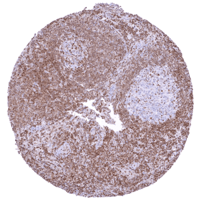
Recombinant Rabbit monoclonal / IgG 1:100 – 1:200 Research Use Only Nuclear and cytoplasmic Human HMV3970 cyclin dependent kinase inhibitor 1B , CDKN4 , KIP1 , MEN1B , MEN4 , P27KIP1 Tonsil: A strong nuclear p27 staining should be seen in a small subset of germinal centre cells and in a large fraction of interfollicular lymphocytic cells. Tonsil: p27 staining should be completely absent in most germinal centre cells. p27/kip1 is a cyclin dependent kinase (Cdk) inhibitor which is coded by the CDKN1B gene at chromosome 12p13. The protein consists of 198 amino acids. Because of its lack of a stable tertiary structure it is classified as an intrinsically disordered protein. The best known role of p27 is its downregulation of cell proliferation through inhibition of most cyclin-Cdk complexes which are needed for cell cycle progression by direct binding to both cyclins and Cdks. p27 includes a large kinase inhibitory domain (KID) containing a cyclin-binding (D1), a Cdk-binding (D2), and a linker subdomain to connect D1 and D2, as well as a nuclear export signal (NES) at aa 32-46. P27 exerts its inhibiting role by inserting D2 into the catalytic center of the Cdks where it competes with ATP and blocks the transfer of phosphate to its substrates. Known transcription factors that activate the cellular production of p27 include KHR-L1, AFX, FOXO, Sp1, E2F1, and menin while c-myc, Id3 and Ap1 repress p27 production. Posttranslational processes which also determine the subcellular localization of p27 may be even more important for the regulation of p27 function than expression alone. They also determine the subcellular localization of p27. Phosphorylation and acetylation of p27 at specific sites can trigger its intranuclear degradation or export into the cytoplasm. Apart from cell cycle regulation, p27 has a role in the regulation of cytoskeleton reorganizationcell migration, apoptosis, autophagy,and gene expression. p27 deregulation such as decreased nuclear levels or cytoplasmic localization are thought to play a role in several types of disease including cancer and neurodegeneration. p27/kip1 is a cyclin dependent kinase (Cdk) inhibitor which is coded by the CDKN1B gene at chromosome 12p13. The protein consists of 198 amino acids. Because of its lack of a stable tertiary structure it is classified as an intrinsically disordered protein. The best known role of p27 is its downregulation of cell proliferation through inhibition of most cyclin-Cdk complexes which are needed for cell cycle progression by direct binding to both cyclins and Cdks. p27 includes a large kinase inhibitory domain (KID) containing a cyclin-binding (D1), a Cdk-binding (D2), and a linker subdomain to connect D1 and D2, as well as a nuclear export signal (NES) at aa 32-46. P27 exerts its inhibiting role by inserting D2 into the catalytic center of the Cdks where it competes with ATP and blocks the transfer of phosphate to its substrates. Known transcription factors that activate the cellular production of p27 include KHR-L1, AFX, FOXO, Sp1, E2F1, and menin while c-myc, Id3 and Ap1 repress p27 prod... p27 staining of at least a subset of nuclei occurs in all organs. Nuclear p27 staining is especially abundant in muscle cells, ovarian stroma, lymphocytes, lung and the brain. In various tissues, p27 expression is more prominent in cell types with a low proliferative activity than in those with high rate of cell division. In lymph nodes, for example, germinal centres contain fewer p27 positive cells than the interfollicular space. Images describing the p27 staining pattern in normal tissues obtained by the antibody HMV3970 are shown in our “Normal Tissue Gallery” . Brain Cerebrum Neurons are mostly negative in the grey matter while there are many p27 positive cells in the white matter. Cerebellum Strong nuclear p27 positivity of most granular cells while Purkinje cells and most cells of the molecular layer are p27 negative. In the white matter there are many strongly p27 positive cells. Endocrine Tissues Thyroid Weak to moderate nuclear p27 positivity of most follicular cells. Parathy... p27 expression – at variable levels – has been described to occur in virtually all cancer types. Reduced expression has been linked to unfavorable tumor features or poor prognosis in many publications. The TCGA findings on p27 RNA expression in different tumor categories have been summarized in the Human Protein Atlas. Prostatic adenocarcinoma (Gleason
4+4=8) with only weak p27 staining of
tumor cells while p27 staining is much
more intense in non-neoplastic
(atrophic) prostate glands. Esophageal adenocarcinoma with moderate to
strong, predominantly nuclear p27 staining of tumor
cells. p27 negative testicular seminoma with strong
p27 staining of inflammatory cells and other
stroma cells. Cancer tissue gallery IHC users have different preferences on how the stains should look like. Some prefer high staining intensity of the target stain and even accept some background. Others favor absolute specificity and lighter target stains. Factors that invariably lead to more intense staining include higher concentration of the antibody and visualization tools, longer incubation time, higher temperature during incubation, higher temperature and longer duration of the heat induced epitope retrieval (slide pretreatment). The impact of the pH during slide pretreatment has variable effects and depends on the antibody and the target protein. All images and data shown here and in on image galleries are obtained by the manual protocol described below. Other protocols resulting in equivalent staining are described as well. Manual protocol Freshly cut sections should be used (less than 10 days between cutting and staining). Heat-induced antigen retrieval for 5 minutes in an autoclave at 121°C in pH9 Target Retr... The prognostic role of p27 alterations needs to be further investigated in cancer. The role of p27 in cytoskeletal dynamics, cell migration, apoptosis, and autophagy needs to be further explored. The role of p27 as a transcriptional regulator awaits further exploration. The role of p27 in neurodegeneration has not been sufficiently clarified. There are two ways how the specificity of antibodies can be documented for immunohistochemistry on formalin fixed tissues. These are: 1. Comparison with a second independent method for target expression measurement across a large number of different tissue types (orthogonal strategy), and 2. Comparison with one or several independent antibodies for the same target and showing that all positive staining results are also seen with other antibodies for the same target (independent antibody strategy). Orthogonal validation: For the antibody HMV3970 specificity is in line with data from three independent RNA screening studies, including the Human Protein Atlas (HPA) RNA-seq tissue dataset, the FANTOM5 project, and the Genotype-Tissue Expression (GTEx) project, which are all summarized in the Human Protein Atlas (Tissue expression p27) . In agreement with HMV3970 immunostaining data, high p27 RNA expression occurs in all normal tissues. However, it must be understood that orthogonal validati...