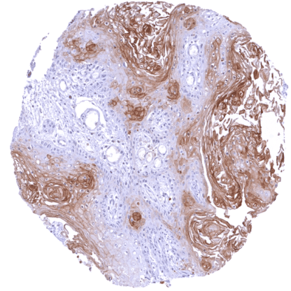
PAI2 (HMV330)
Recombinant Rabbit monoclonal / IgG 1:100 – 1:200 Research Use Only Nuclear Human HMV330 serpin family B member 2,HsT1201,PAI,PAI-2,PAI2,PLANH2 Tonsil: A strong cytoplasmic and nuclear PAI2 staining must be seen in a large fraction of squamous epithelial cells. Tonsil: Lymphocytes must be PAI2 negative (few monocytes/macrophages may show PAI2 staining). PAI2 is an inhibitor of fibrinolysis with a putative role in cancer biology. Plasminogen activator inhibitor-2 (placental PAI, SerpinB2, PAI-2) is a serine protease inhibitor of the serpin superfamily coded by the SerpinB2 gene on chromosome 18.q22.1. It acts as a coagulation factor that irreversibly inactivates tissue plasminogen activator and urokinase. PAI2 exists as a 60-kDa extracellular glycosylated form and a 43-kDa intracellular form. PAI2 protein is normally not detectable in adult plasma. The protein is produced at large quantities in the placenta which explains that PAI2 is detectable in blood only during pregnancy. PAI2 may thus contribute to the increased rate of thrombosis during pregnancy. PAI2 can bind to multiple intracellular and extracellular proteins. For example, it was suggested that PAI2 may activate p53 and stabilize p21. Macrophage derived PAI2 plays a role in inflammatory responses and infections, potentially in downregulating T cells that secrete IgG2c and interferon type II. Although glycosylated extracellular PAI2 regulates fibrinol... Images describing the PAI2 staining pattern in normal tissues obtained by the antibody HMV330 are shown in our “ Normal Tissue Gallery ”. Brain Cerebrum Negative. Cerebellum Negative. Endocrine Tissues Thyroid Negative. Parathyroid Negative. Adrenal gland Negative. Pituitary gland Negative. Respiratory system Respiratory epithelium Negative. Lung Negative. Gastrointestinal Tract Salivary glands Moderate to strong cytoplasmic and nuclear PAI2 staining of epithelial cells from excretory ducts (not in all samples). Esophagus Weak cytoplasmic and nuclear PAI2 staining of top 2/3 cell layers of squamous epithelium (basal and suprabasal layers are PAI2 negative). Stomach Negative. Duodenum Negative. Small intestine Negative. Appendix Negative. Colon Negative. Rectum Negative. Liver Negative. Gallbladder Negative. Pancreas Moderate to strong cytoplasmic and nuclear PAI2 staining of a small subset of epithelial cells. Genitourinary Kidney Negative. Urothelium Negative. Male genital Prostate Ne... PAI2 is most often expressed in squamous cell carcinomas from various sites and in urothelial carcinomas, but PAI2 expression can also occur in tumors of various other organs. The TCGA findings on PAI2 RNA expression in different tumor categories have been summarized in the Human Protein Atlas. Esophageal squamous cell carcinoma with moderate to strong, cytoplasmic and nuclear PAI2 staining of most tumor cells. Oral cavity squamous cell carcinoma with weak to moderate, predominantly cytoplasmic PAI2 staining of most tumor cells. Papillary renal cell carcinoma lacking PAI2 staining in tumor cells. PAI2 positivity of few intratumoral macrophages. Cancer tissue gallery No data available at the moment IHC users have different preferences on how the stains should look like. Some prefer high staining intensity of the target stain and even accept some background. Others favor absolute specificity and lighter target stains. Factors that invariably lead to more intense staining include higher concentration of the antibody and visualization tools, longer incubation time, higher temperature during incubation, higher temperature and longer duration of the heat induced epitope retrieval (slide pretreatment). The impact of the pH during slide pretreatment has variable effects and depends on the antibody and the target protein. All images and data shown here and in our image galleries are obtained by the manual protocol described below. Other protocols resulting in equivalent staining are described as well. Manual protocol Freshly cut sections should be used (less than 10 days between cutting and staining). Heat-induced antigen retrieval for 5 minutes in an autoclave at 121°C in pH 7,8 Target ... The diagnostic and prognostic relevance of PAI2 IHC in tumors and in preneoplastic disease is unresolved. The function/role of intracellular PAI2 has not yet been identified. The role of PAI2 in cancer is unclear. There are two ways how the specificity of antibodies can be documented for immunohistochemistry on formalin fixed tissues. These are: 1. Comparison with a second independent method for target expression measurement across a large number of different tissue types (orthogonal strategy), and 2. Comparison with one or several independent antibodies for the same target and showing that all positive staining results are also seen with other antibodies for the same target (independent antibody strategy). Orthogonal validation: For the antibody HMV330 specificity is supported by the strong concordance with RNA expression data derived from three independent RNA screening studies, including the Human Protein Atlas (HPA) RNA-seq tissue dataset, the FANTOM5 project, and the Genotype-Tissue Expression (GTEx) project, which are all summarized in the Human Protein Atlas (Tissue expression PAI2) . In agreement with these data, PAI2 immunostaining was observed in all tissues covered by squamous epithel...