PARP1 (HMV334)
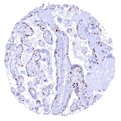
Recombinant Rabbit monoclonal / IgG 1:100 – 1:200 Research Use Only Nuclear Human HMV334 poly(ADP-ribose) polymerase 1 , ADPRT , ADPRT 1 , ADPRT1 , ARTD1 , PARP , PARP-1 , PPOL , pADPRT-1 Placenta: A strong PARP1 positivity should be seen in all cell types except syncytiotrophoblast. Placenta: PARP1 positivity should be absent in the syncytiotrophoblast. PARP1 is a Pleitropic gene with a critical role in DNA repair. Poly(ADP-ribose) polymerase 1 (PARP1), is a 113 kDa nuclear protein coded by the PARP1 gene on chromosome 1q42.12. PARP1 is the first detected and most abundant member of the PARP superfamily. It modulates the activity of DNA binding proteins by catalyzing their Poly(ADP-ribosyl)ation (PARylation), a post-translational modification affecting the conformation and function of affected proteins. PARP1 impacts several DNA repair processes including the pathways of nucleotide excision repair, non-homologous end joining, microhomology-mediated end joining, homologous recombinational repair, and DNA mismatch repair. PARP1 detects DNA damage and then modulates repair efficiency by ADP-ribosylation of histones, subsequent decompaction of chromatin structure, and through interaction with and modification of multiple DNA repair factors. PARP1 and PARylation also play a role in a wide range of further cellular processes such as cell death, chromatin remodeling, inflammatory response and gene tran... Images describing the PARP1 staining pattern in normal tissues obtained by the antibody HMV334 are shown in our “ Normal Tissue Gallery ”. Brain Cerebrum In epithelial cells, PARP1 staining is strongest in the crypts. Cerebellum In epithelial cells, PARP1 staining is strongest in the crypts. Endocrine Tissues Thyroid In epithelial cells, PARP1 staining is strongest in the crypts. Parathyroid In epithelial cells, PARP1 staining is strongest in the crypts. Adrenal gland In epithelial cells, PARP1 staining is strongest in the crypts. Pituitary gland In epithelial cells, PARP1 staining is strongest in the crypts. Respiratory system Respiratory epithelium In epithelial cells, PARP1 staining is strongest in the crypts. Lung In epithelial cells, PARP1 staining is strongest in the crypts. Gastrointestinal Tract Salivary glands In epithelial cells, PARP1 staining is strongest in the crypts. Esophagus Strong PARP1 staining of most squamous epithelial cells. The staining intensity decreases somew... A positive PARP1 immunostaining (intensity may vary) is usually seen in tumor cells of virtually all cancer types as well as in tumor associated stromal and inflammatory cells. The TCGA findings on PARP1 RNA expression in different tumor categories have been summarized in the Human Protein Atlas. Warthin tumor of a salivary gland with a weak
to moderate PARP1 staining of tumor cells
and a markedly more intense staining of
associated lymphocytes. Invasive breast cancer of no special type
(NST) with only a weak to moderate PARP1
staining of tumor cells while the staining is
more intense in tumor-associated
lymphocytes. Endometroid ovarian carcinoma with
intense PARP1 positivity of tumor cells.
The PARP1 staining intensity is
markedly weaker in stromal cells. Cancer tissue gallery No data available at the moment IHC users have different preferences on how the stains should look like. Some prefer high staining intensity of the target stain and even accept some background. Others favor absolute specificity and lighter target stains. Factors that invariably lead to more intense staining include higher concentration of the antibody and visualization tools, longer incubation time, higher temperature during incubation, higher temperature and longer duration of the heat induced epitope retrieval (slide pretreatment). The impact of the pH during slide pretreatment has variable effects and depends on the antibody and the target protein. All images and data shown here and in our image galleries are obtained by the manual protocol described below. Other protocols resulting in equivalent staining are described as well. Manual protocol Freshly cut sections should be used (less than 10 days between cutting and staining). Heat-induced antigen retrieval for 5 minutes in an autoclave at 121°C in pH 7,8 Target ... The clinical significance (prognostic/predictive) of PARP1 expression levels in cancer is unknown. The prevalence of PARP1 expression loss in cancer and its clinical significance is unknown. The role of PARP1 in disease awaits further investigation. There are two ways how the specificity of antibodies can be documented for immunohistochemistry on formalin fixed tissues. These are: 1. Comparison with a second independent method for target expression measurement across a large number of different tissue types (orthogonal strategy), and 2. Comparison with one or several independent antibodies for the same target and showing that all positive staining results are also seen with other antibodies for the same target (independent antibody strategy). Orthogonal validation: For the antibody HMV334 specificity is in line with data from three independent RNA screening studies, including the Human Protein Atlas (HPA) RNA-seq tissue dataset, the FANTOM5 project, and the Genotype-Tissue Expression (GTEx) project, which are all summarized in the Human Protein Atlas (Tissue expression PARP1) . In agreement with HMV334 immunostaining data, RNA expression predominated in the bone marrow and lymphoid tissues and it was comparably low in the testis....