PLAP (MSVA-350R)
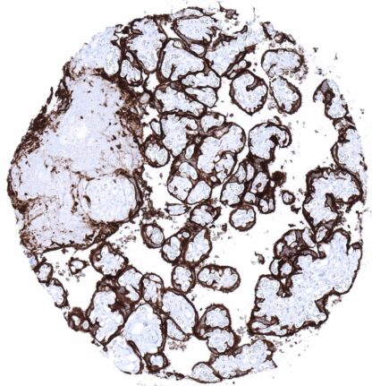
Recombinant Rabbit monoclonal / IgG 1:100 – 1:200 Research Use Only Cell Surface and Cytoplasmic Human MSVA-350R Alkaline phosphatase placental type; Alkaline phosphatase Regan Isozyme; ALP; Alp1; ALPP; Germ-cell alkaline phosphatase; nagao Isozyme; PALP; Placental alkaline phosphatase 1; placental heat-stable alkaline phosphatase; PLAP-1; PLAP1 Placenta (mature): A strong membranous and cytoplasmic staining of cytotrophoblast and syncytiotrophoblast must be seen. Appendix: PLAP immunostaining must be absent in all structures. PLAP is a marker for germ cell tumors which can also be expressed in various types of adenocarcinomas. Placental alkaline phosphatase (PLAP) is a 58 KDa enzyme encoded by the ALPP gene at the human chromosome 2q37.1. The highly polymorphic ALPP gene is expressed in 18 different allelic variants which, however, all share the same function. The transition from the pre-mature to the mature form of the enzyme involves cleavage of the pro-peptide portion spanning amino acids 507-535. Mature PLAP is bound to the plasma membrane. Homodimerization of two PLAP molecules is required to form the catalytic center. The physiological role of PLAP is still unknow, but it might aid in guiding migratory cells and transport specific molecules through the plasma membrane. PLAP becomes expressed in the maturing placenta with steadily raising levels during pregnancy. Three of the different PLAP isoforms have been attributed to to early, mid-term and term placenta. [1] [1] Reiswich et al Reiswich et al “ Pattern of placental alkaline phosphatase (PLAP) expression in human tumors: a tissue microarray study on... PLAP staining pattern in Normal Tissues with antibody MSVA-350R (images are shown in our “Normal Tissue Gallery”) Brain Cerebrum Negative. Cerebellum Negative. Endocrine Tissues Thyroid Negative. Parathyroid Negative. Adrenal gland Negative. Pituitary gland Negative. Respiratory system Respiratory epithelium Negative. Lung Focal membranous PLAP staining of apical membranes of pneumocytes. Gastrointestinal Tract Salivary glands Negative. Esophagus Negative. Stomach Negative. Colon Negative. Duodenum Negative. Rectum Negative. Small intestine Negative. Liver Negative. Gallbladder Negative. Pancreas Negative. Genitourinary Kidney Negative. Urothelium Negative. Male genital Prostate Negative. Seminal vesicles Negative. Testis Negative. Epididymis Negative. Female genital Breast Negative. Uterus, ectocervix Negative. Uterus endocervix A weak PLAP staining can be seen at the surface apical membrane of epithelial cells (not in all samples). Uterus, endometrium A weak PLAP staining can be seen... PLAP is consistently expressed at high levels in seminoma, dysgerminoma, germinoma, intratubular germ cell neoplasia, and hydatidiform moles. PLAP is – usually at lower levels – also often positive in embryonal carcinoma, yolk sack carcinomas, and choriocarcinoma. PLAP is usually absent in spermatocytic seminoma. Detailed data on PLAP staining by MSVA-350R obtained from an analysis of 12,381 tumors from 131 different tumor types and subtypes have recently been published by Reiswich et al “ Pattern of placental alkaline phosphatase (PLAP) expression in human tumors: a tissue microarray study on 12,381 tumors “ The authors found a moderate to strong PLAP positivity in seminoma (96%), embryonal carcinoma (85%), yolk sac tumors of the testis (56%), endometrioid carcinoma of the endometrium (28%) and the ovary (20%), gastric adenocarcinoma (22%), serous carcinoma of the ovary (17%) and the uterus (11%), adenocarcinoma of the ampulla of Vater (15%), carcinosarcoma of the ovary (11%) and the ... PLAP (MSVA-350R) publication summary: Relevant publication: Reiswich et al. “ Pattern of placental alkaline phosphatase (PLAP) expression in human tumors: a tissue microarray study on 12,381 tumors “. Published in the journal of Pathology: Clinical Research, August 7th 2021 A total of 12,381 tumors were analyzed from 131 different tumor categories by using the following protocol: Heat-induced antigen retrieval for 5 minutes in an autoclave at 121°C in pH9 Target Retrieval Solution buffer. MSVA-350R at a dilution of 1:150 at 37°C for 60 minutes. Visualization of bound antibody by the EnVision Kit (Dako, Agilent). This protocol was also used for all stainings depicted in our tumor and normal tissue galleries. At least one case with a positive PLAP immunostaining was seen in 48 (36.6%) and at least one case with a strong PLAP immunostaining was seen in 22 (16.8%) of 131 tumor categories. The distribution of positive staining results is shown “organ-systematic” and in a “ranking order” fi... IHC users have different preferences on how the stains should look like. Some prefer high staining intensity of the target stain and even accept some background. Others favor absolute specificity and lighter target stains. Factors that invariably lead to more intense staining include higher concentration of the antibody and visualization tools, longer incubation time, higher temperature during incubation, higher temperature and longer duration of the heat induced epitope retrieval (slide pretreatment). The impact of the pH during slide pretreatment has variable effects and depends on the antibody and the target protein. Accordingly, multiple different protocols can generate identical staining results. All images and data shown here and in our image galleries are obtained by the manual protocol described below. Other protocols resulting in equivalent staining are described as well. Manual protocol Freshly cut sections should be used (less than 10 days between cutting and staining). Heat... The clinical significance of PLAP expression in non-germ cell neoplasms and the function of PLAP in these tumors needs to be investigated. Specificity of MSVA-350R is documented by strong positive staining in cell types that are documented to express PLAP such as placenta tissue and to a much lesser extent cervical/endometrium/fallopian tube and absence of staining in all other tissues including tissues notorious for non-specific IHC background such as kidney, colonic mucosa, and epidermis. In a recent publication, the specificity of MSVA-350R was demonstrated in a comparison vs. another independent antibody. All normal tissues structures stained by MSVA-350R were also detected with the second independent antibody by Reiswich et al “ Pattern of placental alkaline phosphatase (PLAP) expression in human tumors: a tissue microarray study on 12,381 tumors “ . Normal tissue gallery