PSA (MSVA-603R)
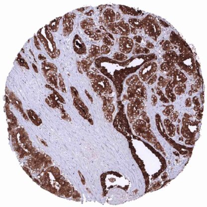
Recombinant Rabbit monoclonal / IgG 1:100 – 1:200 Research Use Only Cytoplasmic Human MSVA-603R Antigen, prostate-specific (APS), Gamma-seminoprotein, hK3, Kallikrein related peptidase 3, Kallikrein-3 (KLK3), KLK2A1, P-30 antigen, Semenogelase, Seminin Prostate: The epithelial cells of the prostate glands must show a strong cytoplasmic staining. Due to leakage of the antigen in the vicinity of the prostate glands, adjacent stroma cells may display a weak to moderate staining reaction. Colon: PSA immunostaining should be absent in all cells. PSA is a marker for prostatic epithelial tissue and prostate cancer. Prostate specific antigen (PSA) is a proteinase coded by the KLK3 gene at 19q13.33. PSA is the most relevant protein for the management of men with suspected or diagnosed and treated prostate cancer. PSA is exclusively produced in prostate epithelial cells. It is secreted to the seminal fluid and plays a role for its liquefaction. Only minor quantities of PSA reach the bloodstream. The serum PSA level is largely proportionate to the quantity of prostate epithelial cells in the body. An increased serum PSA level is the most common cause for prostate cancer suspicion and subsequent prostate biopsy. In men with diagnosed prostate cancer, serum PSA analysis is the most commonly used parameter to monitor disease progression, disease recurrence, and response to therapy. Among normal tissues PSA immunostaining is only seen in prostatic epithelial cells. PSA staining is absent in other normal tissues including aorta, heart, skeletal muscle, smooth muscle, myometrium, ovary (including corpus luteum and follicular cysts), fat, squamous epithelia irrespective of their origin, anal canal transitional epithelium, urothelium, stomach, duodenum, small intestine, appendix, colon, rectum, gallbladder, bronchus, paranasal sinus, lymphatic tissues, node, spleen, thymus, tonsil, liver, pancreas, parotid gland, submandibular gland, sublingual gland, Brunner gland, kidney, seminal vesicle, epididymis, testis, lung, breast, endocervix, endometrium, fallopian tube, placenta, adrenal gland, parathyroid gland, thyroid, cerebellum, cerebrum, and the pituitary gland. The findings described above are this consistent with the RNA data described in the Human Protein Atlas (Tissue expression PSA) Positive control = Prostate: The epithelial cells of the prostate glands must sh... PSA immunostaining is almost completely restricted to prostate cancers. PSA expression is seen in >98% of prostatic adenocarcinomas at the time of initial diagnosis. Advanced high grade prostate cancers can gradually lose PSA expression during tumor progression, however. The TCGA findings on PSA RNA expression in different tumor categories have been summarized in the Human Protein Atlas. Adenocarcinoma (Gleason 3+3=6) with intense PSA immunostaining of tumor cells. PSA negative endometroid carcinoma. Adenocarcinoma (Gleason 5+5=10) with moderate to strong PSA immunostaining of tumor cells. Cancer tissue gallery No data available at the moment IHC users have different preferences on how the stains should look like. Some prefer high staining intensity of the target stain and even accept some background. Others favor absolute specificity and lighter target stains. Factors that invariably lead to more intense staining include higher concentration of the antibody and visualization tools, longer incubation time, higher temperature during incubation, higher temperature and longer duration of the heat induced epitope retrieval (slide pretreatment). The impact of the pH during slide pretreatment has variable effects and depends on the antibody and the target protein. All images and data shown here and in our image galleries are obtained by the manual protocol described below. Other protocols resulting in equivalent staining are described as well. Manual protocol Freshly cut sections should be used (less than 10 days between cutting and staining). Heat-induced antigen retrieval for 5 minutes in an autoclave at 121°C in pH 7,8 Target ... The clinical significance of reduced expression of PSA in prostate cancers should be further investigated. The level of PSA expression (thought to be related to patient prognosis) might constitute an important parameter for multiparametric prostate cancer prognosis tests There are two ways how the specificity of antibodies can be documented for immunohistochemistry on formalin fixed tissues. These are: 1. Comparison with a second independent method for target expression measurement across a large number of different tissue types (orthogonal strategy), and 2. Comparison with one or several independent antibodies for the same target and showing that all positive staining results are also seen with other antibodies for the same target (independent antibody strategy). Orthogonal validation: For the antibody MSVA-603R specificity is suggested by the perfect concordance of the immunostaining data with data from three independent RNA screening studies, including the Human Protein Atlas (HPA) RNA-seq tissue dataset, the FANTOM5 project, and the Genotype-Tissue Expression (GTEx) project, which are all summarized in the Human Protein Atlas (Tissue expression PSA) . In these databases, PSA RNA expression was solely expressed in the prostate which also was the onl...