S100P (MSVA-480R)
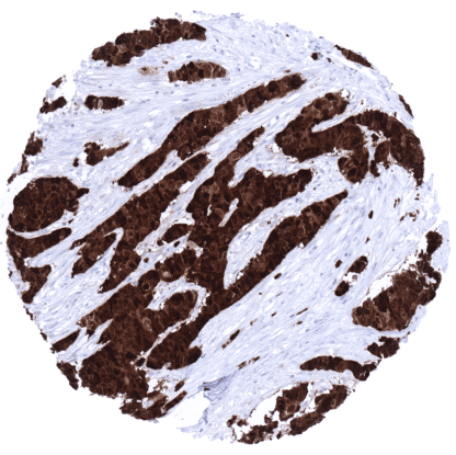
Recombinant Rabbit monoclonal / IgG 1:100-200 Research Use Only Nucleus and cytoplasm Human MSVA-480R Migration inducing gene 9 (MIG9); S100E; S100 calcium binding protein P Urinary bladder: A strong S100P immunostaining should be seen in the urothelium but not in the stroma or muscular cells. Kidney: S100P immunostaining should be completely absent. S100P is expressed in placenta, urothelium, stomach, and respiratory epithelium. S100P is a 10.4 kDa protein , coded by the S100P gene at 4p16. The S100 genes are a group of water soluble low-molecular-weight (9–12 kD) proteins characterized by two calcium-binding sites that have a specific helix-loop-helix (“EF-hand type”) conformation. The “S100” gene name is derived from the fact that these proteins are soluble in 100%. S100P interacts, directly or indirectly, with a number of different proteins, many of which regulate actin cytoskeleton dynamics and extracellular matrix remodeling. Interaction partners for example include Ezrin, IQGAP1, myosin IIA, cathepsin D, and cofilin. Through these interactions, S100P integrates and regulates various signaling pathways and induces a broad range of important functional results. A strong S100P immunostaining is regularly seen in trophoblastic (syncytiotrophoblast more than cytotrophoblast) and chorion cells of the placenta, all cell layers of urothelium (stronger intensity of the top layers than in the basal layers), surface epithelial cells and neck cells (but not of glands) of the stomach, and in granulocytes. A weak to moderate S100P staining can be found in variable fractions of epithelial cells of the colon mucosa (stronger in superficial than in crypt cells), duodenum and jejunum (mainly in goblet cells), transitional epithelium of the anal canal, as well as in mucinous and basal cells of sublingual (but not submandibular) glands. In the tonsil crypts, a weak to moderate S100P staining occurs in the superficial layers of squamous epithelium. In the thymus, a moderate staining of corpuscles of Hassall’s but not of other epithelial cells is seen. S100P can occasionally also occur in several other normal tissues, perhaps due to specific functional changes. ... S100P immunostaining is particularly strong and frequent in urothelial, colorectal and gastrointestinal tumors. Various other tumor entities have been described to express S100P in a fraction of cases. The TCGA findings on S100P RNA expression in different tumor categories have been summarized in the Human Protein Atlas. Muscle-invasive urothelial carcinoma with strong S100P immunostaining of tumor cells. Gastric adenocarcinoma (intestinal type) with strong S100P immunostaining of tumor cells. S100P positive granulocytes are also discernible. Colorectal adenocarcinoma with strong S100P immunostaining of tumor cells. Cancer tissue gallery No data available at the moment IHC users have different preferences on how the stains should look like. Some prefer high staining intensity of the target stain and even accept some background. Others favor absolute specificity and lighter target stains. Factors that invariably lead to more intense staining include higher concentration of the antibody and visualization tools, longer incubation time, higher temperature during incubation, higher temperature and longer duration of the heat induced epitope retrieval (slide pretreatment). The impact of the pH during slide pretreatment has variable effects and depends on the antibody and the target protein. All images and data shown here and in our image galleries are obtained by the manual protocol described below. Other protocols resulting in equivalent staining are described as well. Manual protocol Freshly cut sections should be used (less than 10 days between cutting and staining). Heat-induced antigen retrieval for 5 minutes in an autoclave at 121°C in pH 7,8 Target ... The diagnostic utility of S100 expression analysis should be further investigated in a large cohort of tumors from different entities The diagnostic role of S100P as a parameter of malignancy needs to be investigated. The prognostic role of S100P expression in tumors is not sufficiently analyzed so far. The role of S100P in inflammatory disease should be further evaluated. There are two ways how the specificity of antibodies can be documented for immunohistochemistry on formalin fixed tissues. These are: 1. comparison with a second independent method for target expression measurement across a large number of different tissue types (orthogonal strategy), and 2. Comparison with one or several independent antibodies for the same target and showing that all positive staining results are also seen with other antibodies for the same target (independent antibody strategy). Orthogonal validation: For the antibody MSVA-480R specificity is suggested by the strong concordance of the immunostaining data with data from three independent RNA screening studies, including the Human Protein Atlas (HPA) RNA-seq tissue dataset, the FANTOM5 project, and the Genotype-Tissue Expression (GTEx) project, which are all summarized in the Human Protein Atlas (Tissue expression S100P) . Immunostaining by using MSVA-480R was almost exclusively detected in organs and cell types with d...