SDHA (HMV336)
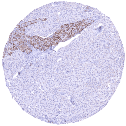
Recombinant Rabbit monoclonal / IgG 1:100 – 1:200 Research Use Only Intracellular Human HMV336 Succinate dehydrogenase complex flavoprotein subunit A , CMD1GG , FP , PGL5 , SDH1 , SDH2 , SDHF Appendix: A strong SDHA staining should be seen in epithelial cells and a weak to moderate staining should occur in lymphocytic cells. SDHA staining should be absent in all cells of a tumor with known SDHA expression loss. SDHA is a critical protein component of the SDH complex. Succinate dehydrogenase complex iron sulfur subunit A (SDHA) is coded by the SDHA gene at chromosome 5p15.33. SDHA forms the succinate dehydrogenase protein complex II together with SDHB, SDHC and SDHD. Within this complex, SDHA is linked to SDHB at the catalytic end of the SDH protein complex which protrudes into the mitochondrial matrix while SDHC and SDHD are hydrophobic and attach the SDH complex to the inner mitochondrial membrane. SDH plays a pivotal role in both the citric acid cycle and the respiratory chain. It catalyzes the conversion of succinate to fumarate in the citric acid cycle downstream of IDH2. A loss of enzyme activity results in accumulation of metabolic intermediates similarly as under hypoxic conditions. Such a “pseudohypoxia” results in HIF-1 pathway activation and may induce a metabolic shift toward aerobic glycolysis . SDHA germline mutations can cause pheochromocytoma, paraganglioma, gastrointestinal stromal tumor (GIST), pituitary adenoma and renal cell carc... Images describing the SDHA staining pattern in normal tissues obtained by the antibody HMV336 are shown in our “ Normal Tissue Gallery ”. Brain Cerebrum Rather weak granular cytoplasmic SDHA staining of cells. Cerebellum Rather weak granular cytoplasmic SDHA staining of cells. Endocrine Tissues Thyroid Weak granular cytoplasmic SDHA staining of epithelial cells. Parathyroid Weak to strong granular cytoplasmic SDHA staining of epithelial cells. Marked heterogeneity between cells and cell groups. Adrenal gland Strong granular cytoplasmic SDHA staining of epithelial cells. Pituitary gland Strong granular cytoplasmic SDHA staining of epithelial cells. Moderate granular cytoplasmic SDHA staining of pituicytes and fibres. Respiratory system Respiratory epithelium Marked granular cytoplasmic SDHA staining of epithelial cells, predominately in the sub-apical compartment. Lung SDHA staining is largely absent in alveocytes. SDHA staining is prominent in macrophages and bronchiole.. Gastrointesti... A variable level of SDHA expression occurs in virtually all tumors except those with an SDH deficiency. According to RNA data of the TCGA database, the level of SDHA expression is highly prognostic in renal cell carcinomas. The TCGA findings on SDHA RNA expression in different tumor categories have been summarized in the Human Protein Atlas. Urinary bladder showing an SDHA
negative muscle-invasive urothelial
carcinoma. SDHA staining is intense in
tumor infiltrating inflammatory cells. Pulmonaly adenocarcinoma showing a
strong SDHA staining of all tumor cells. Prostatic adenocarcinoma (Gleason 5+5=10)
with only a faint positivity of tumor cells
while SDHA staining is markedly more
intense in two entrapped normal glands. Cancer tissue gallery No data available at the moment IHC users have different preferences on how the stains should look like. Some prefer high staining intensity of the target stain and even accept some background. Others favor absolute specificity and lighter target stains. Factors that invariably lead to more intense staining include higher concentration of the antibody and visualization tools, longer incubation time, higher temperature during incubation, higher temperature and longer duration of the heat induced epitope retrieval (slide pretreatment). The impact of the pH during slide pretreatment has variable effects and depends on the antibody and the target protein. All images and data shown here and in our image galleries are obtained by the manual protocol described below. Other protocols resulting in equivalent staining are described as well. Manual protocol Freshly cut sections should be used (less than 10 days between cutting and staining). Heat-induced antigen retrieval for 5 minutes in an autoclave at 121°C in pH 7,8 Target ... The prognostic relevance of SDHA expression in tumors and in preneoplastic disease needs to be investigated. The predictive relevance of SDHA expression in tumors is unknown. The role of SDHA expression levels and of SDHA mutation in non-neoplastic disease is unknown. There are two ways how the specificity of antibodies can be documented for immunohistochemistry on formalin fixed tissues. These are: 1. Comparison with a second independent method for target expression measurement across a large number of different tissue types (orthogonal strategy), and 2. Comparison with one or several independent antibodies for the same target and showing that all positive staining results are also seen with other antibodies for the same target (independent antibody strategy). Orthogonal validation: For the antibody HMV336 specificity is supported by the good concordance of the immunostaining data with data from three independent RNA screening studies, including the Human Protein Atlas (HPA) RNA-seq tissue dataset, the FANTOM5 project, and the Genotype-Tissue Expression (GTEx) project, which are all summarized in the Human Protein Atlas (Tissue expression SDHA) . SDHA positivity by HMV336 is particularly strong in heart and skeletal muscle, liver, kidney, parathyro...