SOX9 (MSVA-709R)
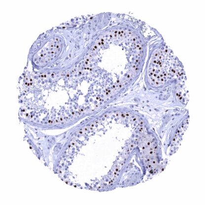
Recombinant Rabbit monoclonal / IgG 1:50 – 1:75 Research Use Only Nucleus Human MSVA-709R Campomelic Dysplasia Autosomal Sex Reversal (CMD1); SRA1; SRXX2; SRY (sex determining region Y) box 9; SRY related HMG box gene 9; Transcription factor SOX9 Colon: A strong SOX9 immunostaining should be seen in epithelial cells of the crypt base. Colon: SOX9 immunostaining should be absent in all non-epithelial cell types (except few cells in lymphoid germinal centres – if present). SOX-9 is an kDa transcription factor protein coded by the SOX-9 gene on chromosome 17q24. SOX-9 specifically recognizes the sequence CCTTGAG. SOX-9 is a regulatory element for the development of cells and tissues. It shows well-defined temporal and spatial expression patterns that differ between particular cell types and tissues and thus exerts a role in cell lineage restriction and terminal differentiation. For example, SOX-9 plays a pivotal role in differentiation of chondrocytes and in male sexual development. By interacting with multiple other genes, SOX-9 inhibits the creation of a female reproductive system. In several adult ectoderm- and endoderm-derived tissues, SOX-9 expression is retained in stem cell pools. SOX-9 is also believed to play a role in cancer stem cells. SOX9 staining pattern in Normal Tissues with antibody MSVA-709R (Images shown in our “Normal Tissue Gallery”) Brain Cerebrum Negative*. Cerebellum Negative*. Endocrine Tissues Thyroid Moderate to strong SOX9 positivity of follicular cells. Parathyroid Negative. Adrenal gland Few interspersed cells in the cortex showing weak to moderate SOX9 positivity. Pituitary gland Respiratory system Respiratory epithelium Moderate to strong SOX9 positivity of most epithelial cells. Lung Pneumocytes and alveolar cells are SOX9 negative. Gastrointestinal Tract Salivary glands Moderate to strong nuclear SOX9 staining of all epithelial cell types. Esophagus Weak to moderate nuclear SOX9 staining of the bottom 2/3 of squamous epithelium. Stomach Moderate to strong nuclear SOX9 staining of surface epithelial cells, while the glandular cells are either SOX9 negative or only show weak positivity. Duodenum Strong nuclear SOX9 staining of crypt epithelial cells. The SOX9 staining intensity markedly decreases... Tumors with variable levels of SOX9 immunostaining can be seen within a variety of different tumor entities. The TCGA findings on SOX9 RNA expression in different tumor categories have been summarized in the Human Protein Atlas. Uterus, cervix – Adenocarcinoma with strong SOX9 positivity of 100% of tumor cells Stomach – Gastric adenocarcinoma (diffuse type) with intense SOX9 immunostaining of tumor cells Salivary gland – Warthin tumor with strong SOX9 staining of most epithelial tumor cells Cancer tissue gallery No data available at the moment IHC users have different preferences on how the stains should look like. Some prefer high staining intensity of the target stain and even accept some background. Others favor absolute specificity and lighter target stains. Factors that invariably lead to more intense staining include higher concentration of the antibody and visualization tools, longer incubation time, higher temperature during incubation, higher temperature and longer duration of the heat induced epitope retrieval (slide pretreatment). The impact of the pH during slide pretreatment has variable effects and depends on the antibody and the target protein. All images and data shown here and in our image galleries are obtained by the manual protocol described below. Other protocols resulting in equivalent staining are described as well. Manual protocol Freshly cut sections should be used (less than 10 days between cutting and staining). Heat-induced antigen retrieval for 5 minutes in an autoclave at 121°C in pH 7,8 Target ... The role of SOX9 is of considerable interest in many different fields such as for example in cancer, regeneration, and stem cell research. The prognostic role of SOX9 expression in cancer is under investigation. There are two ways how the specificity of antibodies can be documented for immunohistochemistry on formalin fixed tissues. These are: 1. comparison with a second independent method for target expression measurement across a large number of different tissue types (orthogonal strategy), and 2. Comparison with one or several independent antibodies for the same target and showing that all positive staining results are also seen with other antibodies for the same target (independent antibody strategy). Orthogonal validation: Data derived from three independent RNA screening studies, including the Human Protein Atlas (HPA) RNA-seq tissue dataset, the FANTOM5 project, and the Genotype-Tissue Expression (GTEx) project, which are all summarized in the Human Protein Atlas (Tissue expression SOX9) suggest that SOX9 expression occurs in a very broad variety of tissues. That SOX9 RNA expression was particularly low or undetectable in the few organs without detectable protein expression (muscles of ...