Cytokeratin 10 (MSVA-610M)
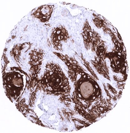
Recombinant Mouse monoclonal / IgG1 1:100 – 1:200 Research Use Only Cytoplasmic Human MSVA-610M BCIE, BIE, EHK, Keratin Type I Cytoskeletal 10, KRT10 Skin: All squamous epithelial cells (except the basal cell layer) should show strong cytoplasmic staining, while the basal cell layer remains KRT10 negative or shows only minimal staining. Colon: All epithelial, inflammatory, stromal and muscle cells must not show any KRT10 staining. Cytokeratin 10 is expressed in keratinizing squamous epithelial cells Cytokeratin 10 (CK10), also termed keratin 10 (KRT10) is a type I acidic high molecular weight keratin protein encoded by the KRT10 gene located 17q21. It dimerizes with the type I keratin 1 and forms intermediate filaments that primarily shape the cytoskeleton of specific epithelial cells. In these cells, KRT10 is part of the cytoskeletal scaffold which contributes to the cell architecture and provides the cells with the ability to withstand mechanical stress. Mutations in CK10 can cause epidermolytic hyperkeratosis or bullous congenital ichthyosiform erythroderma of Brocq. Cytokeratin 10 staining pattern in Normal Tissues with antibody MSVA-610M (images are shown in our “Normal Tissue Gallery”) Brain Cerebrum Negative. Cerebellum Negative. Endocrine Tissues Thyroid Negative. Parathyroid Negative. Adrenal gland Negative. Pituitary gland Negative. Respiratory system Respiratory epithelium Negative. Lung Negative. Gastrointestinal Tract Salivary glands Negative. Esophagus Usually negative. Stomach Negative. Duodenum Negative. Small intestine Negative. Appendix Negative. Colon Negative. Rectum Negative. Liver Negative. Gallbladder Negative. Pancreas Negative. Genitourinary Kidney Negative. Urothelium Negative. Male genital Prostate Negative. Seminal vesicles Negative. Testis Negative. Epididymis Negative. Female genital Breast Negative. Uterus, myometrium Negative. Uterus, ectocervix Usually negative. Uterus endocervix Negative. Uterus, endometrium Negative. Fallopian Tube Negative. Ovary Negative. Placenta early Negative. Placenta mature Negative. Amnion Ne... KRT10 immunostaining almost exclusively occurs in squamous cell carcinomas of various sites of origin. The rate of KRT10 positive cases may vary depending on the histologic tumor grade. Some of the KRT10 positive squamous cell carcinomas show – similar to the situation in normal skin – absence of expression in the basal cell layers. The TCGA findings on Cytokeratin 10 RNA expression in different tumor categories have been summarized in the Human Protein Atlas. Squamous cell carcinoma showing diffuse positivity of 95% of tumor cells. Few peripheral tumor cells are negative or only weakly positive. Neuroendocrine tumor of the pancreas showing a weak to moderate KRT10 immunostaining of most tumor cells. Squamous cell carcinoma of the oral cavity with KRT10 positivity of variable intensity predominantly occurring in the center of tumor cell nests. Cancer tissue gallery Cytokeratin 10 (CK10) (MSVA-610M) publication summary Relevant publication: Uhlig et al. : “Cytokeratin 10 (CK10) expression in cancer: A tissue microarray study on 11,021 tumors” . Published in Ann Diagn Pathol. 2022 Aug 20;60:152029. Epub ahead of print. PMID: 36029589. A total of 9’462 tumors were successfully analyzed from 131 different tumor categories by using the following protocol: Heat-induced antigen retrieval for 5 minutes in an autoclave at 121°C in pH7,8 Target Retrieval Solution buffer. MSVA-610M at a dilution of 1:150 at 37°C for 60 minutes. Visualization of bound antibody by the EnVision Kit (Dako, Agilent). This protocol was also used for all stainings depicted in our tumor and normal tissue galleries. In this study, at least one CK10 positive case was seen in 41 (31,3%) of 131 tumor categories and 18 (13,7%) tumor categories included at least one case with strong positivity. Uhlig et al described the highest CK10 positivity rates in squamous cell carcinomas from vario... IHC users have different preferences on how the stains should look like. Some prefer high staining intensity of the target stain and even accept some background. Others favor absolute specificity and lighter target stains. Factors that invariably lead to more intense staining include higher concentration of the antibody and visualization tools, longer incubation time, higher temperature during incubation, higher temperature and longer duration of the heat induced epitope retrieval (slide pretreatment). The impact of the pH during slide pretreatment has variable effects and depends on the antibody and the target protein. Accordingly, multiple different protocols can generate identical staining results. All images and data shown here and in our image galleries were obtained by the manual protocol described below. Other protocols resulting in equivalent staining are described as well. Manual protocol Freshly cut sections should be used (less than 10 days between cutting and staining). Hea... The diagnostic utility of reduced CK10 expression in the skin is not known. The diagnostic utility of CK10 neo-expression in non-keratinizing squamous epithelium of ectocervix, esophagus, oral cavity and lip is not known. The prognostic role of CK10 expression in squamous cell carcinoma is unknown. Utility of MSVA-610M is documented by a staining pattern that exactly matches the data summarized in the protein atlas. A strong positive KRT10 staining almost exclusively seen in the skin. All other tissues showing weak or occasional KRT10 staining are also squamous epithelia. In addition, all tissues known to not express KRT10 including those notorious for non-specific IHC background such as kidney and colonic mucosa are completely KRT10 negative. Normal tissue gallery