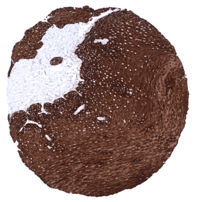
Cytokeratin 15 (MSVA-615M)
Mouse monoclonal / IgG2b 1:50 – 1:100 Research Use Only Cytoplasmic Human MSVA-615M CK15; Cytokeratin 15; K1CO; Ka15; Keratin 15 basic; Keratin 15 beta; Keratin complex 1 acidic gene 15; Keratin type I cytoskeletal 15; KRT15; KRTB; KRTL15; Type I cytoskeletal 15; Type I keratin Ka15 Tonsil: All cells of the surface squamous epithelium and most squamous cells of the crypts should show strong KRT15 staining. Tonsil: All lymphocytes and blood vessels must not show any KRT15 staining. Cytokeratin 15 is physiologically expressed in squamous epithelium and urothelium. Cytokeratin 15 (CK15) also termed keratin 15 (KRT15) is a type I acidic high molecular weight keratin protein encoded by the KRT15 gene located 17q21.2. It dimerizes with the basic type II keratin 4 and forms intermediate filaments that primarily shape the cytoskeleton of specific epithelial cells. In these cells, KRT15 is part of the cytoskeletal scaffold which contributes to the cell architecture and provides the cells with the ability to withstand mechanical stress. KRT15 can be downregulated in activated keratinocytes. A strong KRT15 immunostaining is found in all cell layers of all non-keratinizing and keratinizing squamous epithelium including hair follicles and sebaceous glands, acinar but not myoepithelial cells of the breast, amnion cells of the placenta, urothelium (stronger staining in lower half as compared to upper half), myoepithelial cells and basal cells of excretion ducts of all salivary glands and bronchial glands. In the prostate, epididymis, and the respiratory epithelium, KRT15 immunostaining is strong in all basal cells and occasionally also occurs in luminal epithelial cells. Only few basal cells are KRT15 positive in seminal vesicles. In the thymus, KRT15 is strongly expressed in Hassall’s corpuscles and medullary thymic epithelial cells but weaker in cortical thymic epithelial cells. KRT15 immunostaining can also be seen in the syncytiotrophoblast of the placenta (generally weak and more common in first trimenon than in mature placenta), occasionally in few cells of collecting du... KRT15 immunostaining almost exclusively occurs in squamous cell carcinomas of various sites of origin. At lower frequencies and lower levels, KRT15 expression can also be seen in other tumor types. The TCGA findings on Cytokeratin 15 RNA expression in different tumor categories have been summarized in the Human Protein Atlas. Esophageal squamous cell carcinoma showing strong CK15 immunostaining. Squamous cell carcinoma of the oral cavity with strong CK15 staining. Ovarian Brenner tumor showing moderate Cytokertin 15 staining in tumor cells. Cancer tissue gallery No data available at the moment IHC users have different preferences on how the stains should look like. Some prefer high staining intensity of the target stain and even accept some background. Others favor absolute specificity and lighter target stains. Factors that invariably lead to more intense staining include higher concentration of the antibody and visualization tools, longer incubation time, higher temperature during incubation, higher temperature and longer duration of the heat induced epitope retrieval (slide pretreatment). The impact of the pH during slide pretreatment has variable effects and depends on the antibody and the target protein. Accordingly, multiple different protocols can generate identical staining results. All images and data shown here and in our image galleries are obtained by the manual protocol described below. Other protocols resulting in equivalent staining are described as well. -Manual protocol Freshly cut sections should be used (less than 10 days between cutting and staining). Hea... Studies have suggested that reduced expression in squamous epithelium may be found in case of dysplasia or other pathologic changes. Further investigations on this subject are needed. The prognostic role of KRT15 expression in squamous cell carcinoma is unknown. Specificity of MSVA-615M is documented by a staining pattern that exactly matches the data summarized in the protein atlas. As suggested by the protein atlas data, a strong positive KRT15 staining is seen in all keratinizing and non-keratinizing squamous epithelia. KRT15 staining in specific cell types is also found in other organs where the RNA data of the protein atlas suggest KRT15 expression such as salivary glands, prostate, and respiratory epithelium. In addition, all tissues known to not express KRT15 including those notorious for non-specific IHC background such as kidney and colonic mucosa are completely KRT15 negative. Normal tissue gallery