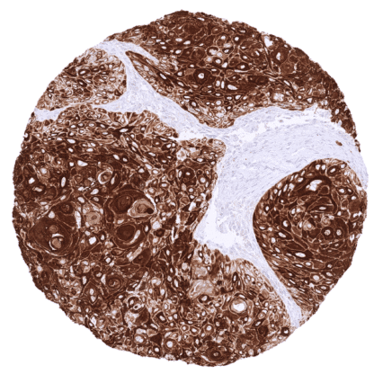
Cytokeratin 17 (MSVA-117R)
Recombinant Rabbit monoclonal / IgG 1:100 – 1:200 Research Use Only Cytoplasmic Human MSVA-117R Keratin-17; KRT17; PCHC1, K17; Keratin Type I Cytoskeletal 17 Prostate: A strong CK17 staining should be seen in basal cells while acinar cells remain negative. Colon: CK17 staining should be absent in all cells. Cytokeratin 17 is Marker for basal cells in many tissues. Keratin 17 (CK17), also termed keratin 17 (KRT17) is an 48 kDa type I cytokeratin coded by the KRT17 gene at 17q21.2. CK17 is part of the cytoskeletal scaffold within epithelial cells, which contributes to the cell architecture and provides the cells with the ability to withstand mechanical stress. It is primarily found in nail beds, sebaceous glands, hair follicles, and other epidermal appendages. CK17 is usually absent in squamous epithelium but is inducible under stressful conditions such as skin injury, viral infections or other inflammatory diseases. Mutations in CK17 are associated with pachyonychia congenita type 2 and steatocystoma multiplex. Images describing the Cytokeratin 17 staining pattern in normal tissues obtained by the antibody MSVA-117R are shown in our “ Normal Tissue Gallery ”. Brain Cerebrum Negative. Cerebellum Negative. Endocrine Tissues Thyroid Negative. Parathyroid Negative. Adrenal gland Negative. Pituitary gland Negative. Respiratory system Respiratory epithelium Strong CK17 staining of basal cells. Lung Negative. Gastrointestinal Tract Salivary glands Strong CK17 staining of basal cells of excretory ducts and of myoepithelial cells. Glandular cells are not stained. Esophagus Few basal cells show weak to moderate staining in some of the samples. Stomach Negative. Colon Negative. Duodenum Negative. Rectum Negative. Small intestine Negative. Liver Negative. Gallbladder Negative. Pancreas Strong CK17 staining of a subset of small excretory ducts while intercalated ducts and large excretory ducts are CK17 negative. Genitourinary Kidney A subset of tubular cells can show strong CK17 positivity in a subset of ... CK17 is primarily expressed in squamous cell carcinomas of various organs of origin and in urothelial carcinomas. CK17 expression may also occur in other tumors. The TCGA findings on Cytokeratin 17 RNA expression in different tumor categories have been summarized in the Human Protein Atlas. Cervical adenocarcinoma with strong CK17 staining of tumor cells Ductal adenocarcinoma of the pancreas with strong CK17 positivity of tumor cells Gastric adenocarcinoma (intestinal type) exhibiting strong CK17 positivity of tumor cells Cancer tissue gallery No data available at the moment IHC users have different preferences on how the stains should look like. Some prefer high staining intensity of the target stain and even accept some background. Others favor absolute specificity and lighter target stains. Factors that invariably lead to more intense staining include higher concentration of the antibody and visualization tools, longer incubation time, higher temperature during incubation, higher temperature and longer duration of the heat induced epitope retrieval (slide pretreatment). The impact of the pH during slide pretreatment has variable effects and depends on the antibody and the target protein. All images and data shown here and in our image galleries are obtained by the manual protocol described below. Other protocols resulting in equivalent staining are described as well. Manual protocol Freshly cut sections should be used (less than 10 days between cutting and staining). Heat-induced antigen retrieval for 5 minutes in an autoclave at 121°C in pH 7,8 Target ... The diagnostic and prognostic relevance of CK17 expression in tumors and in preneoplastic disease needs to be investigated. There are two ways how the specificity of antibodies can be documented for immunohistochemistry on formalin fixed tissues. These are: 1. Comparison with a second independent method for target expression measurement across a large number of different tissue types (orthogonal strategy), and 2. Comparison with one or several independent antibodies for the same target and showing that all positive staining results are also seen with other antibodies for the same target (independent antibody strategy). Orthogonal validation: For the antibody MSVA-117R specificity is suggested by the strong concordance of the immunostaining data with data from three independent RNA screening studies, including the Human Protein Atlas (HPA) RNA-seq tissue dataset, the FANTOM5 project, and the Genotype-Tissue Expression (GTEx) project, which are all summarized in the Human Protein Atlas (Tissue expression Cytokeratin 17 . CK17 positivity by MSVA-117R is detectable in all tissues with documented CK17 RNA expres...