Cytokeratin 18 (MSVA-118R)
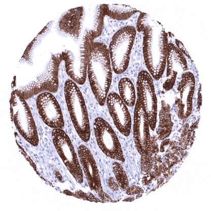
Recombinant Rabbit monoclonal / IgG 1:100 – 1:200 Research Use Only Cytoplasmic Human MSVA-118R Cell Proliferation-inducing Gene 46 Protein; CK18; CYK18Cytokeratin Endo B; K18; Keratin-18; Kerd; KRT18 Liver: At least a weak to moderate, predominantly membranous staining should be seen in virtually all hepatocytes. Appendix: A strong staining should be seen in all epithelial cells. Appendix: Staining should be absent in all non-epithelial cells of the appendix. Cytokeratin 18 (CK18), also termed keratin 18 (KRT18) is an acidic type I keratin protein encoded by the KRT18 gene on 12q13. It forms heteropolymers with his co-expressed complementary type II keratin partner KRT8, which – assembled into keratin filaments – shape the cytoskeleton of many epithelial cell types. As other cytokeratins, KRT18 is part of the cytoskeletal scaffold within epithelial cells, which contributes to the cell architecture and provides the cells with the ability to withstand mechanical stress. CK18 is primarily expressed in single-layered or “simple” epithelial tissues. Beside the important structural function, CK18 was also suggested to play a role in the regulation of cancer associated processes such as apoptosis, cell cycle progression, and cancer-related signaling pathways.[1] Cytokeratin 18 (CK18), also termed keratin 18 (KRT18) is an acidic type I keratin protein encoded by the KRT18 gene on 12q13. It forms heteropolymers with his co-expressed complementary type II keratin partner KRT8, which – assembled into keratin filaments – shape the cytoskeleton of many epithelial cell types. As other cytokeratins, KRT18 is part of the cytoskeletal scaffold within epithelial cells, which contributes to the cell architecture and provides the cells with the ability to withstand mechanical stress. CK18 is primarily expressed in single-layered or “simple” epithelial tissues. Beside the important structural function, CK18 was also suggested to play a role in the regulation of cancer associated processes such as apoptosis, cell cycle progression, and cancer-related signaling pathways. [1] [1] Menz et al. “Diagnostic and prognostic impact of cytokeratin 18 expression in human tumors. a tissue microarray study on 11,952 tumors” Molecular Medicine 27, Article number: 16 (2021... Cytokeratin 18 staining pattern in Normal Tissues with antibody MSVA-118R (images are shown in our “Normal Tissue Gallery”) Brain Cerebrum Negative. Cerebellum Negative. Endocrine Tissues Thyroid Moderate to strong staining of follicular cells. Parathyroid Moderate staining of all epithelial cells. Adrenal gland Negative. Pituitary gland Strong staining of epithelial cells in the adenohypophysis. Respiratory system Respiratory epithelium Strong CK18 positivity of epithelial cells but staining may be weaker in basal cells. Lung Strong CK18 positivity of all pneumocytes. Gastrointestinal Tract Salivary glands CK18 staining is strong in serous and mucinous cells but is generally weaker in excretory ducts. Individual excretory ducts also show weak or absent staining. Esophagus Negative. Stomach Strong CK18 positivity of most epithelial cells. Staining is weak or absent in parietal cells of the corpus. Duodenum Strong CK18 positivity of all epithelial cells. Small intestine Strong CK18 posi... KRT18 expression is detectable in many different tumor types. High level KRT18 expression commonly occurs in many epithelial tumors, mainly in those derived from KRT18 positive normal tissues. Tumor types which are almost always KRT18 positive include for example adenocarcinomas of the colorectum, stomach, lung, prostate, pancreas, thyroid as well as ovarian cancer. Tumor types that are mostly KRT18 negative for example include squamous cell carcinomas from various sites of origin, seminomas, and soft tissue tumors. Loss of KRT18 expression in cancers derived from KRT18 expressing precursor cells and upregulation or neoexpression of KRT18 in neoplasias derived from KRT18 negative precursor cells is often linked to unfavorable tumor phenotype and poor patient outcome. Detailed data on CK18/KRT18 staining by MSV-118R obtained from an analysis of 11,952 tumors from 115 different tumor types and subtypes have recently been published by Menz et al. “Diagnostic and prognostic impact of cyto... Cytokeratin 18 (MSVA-118R) publication summary (MSVA-118R is the recombinant version of the published antibody clone MSVA-118M) Relevant publication: Menz et al. “Diagnostic and prognostic impact of cytokeratin 18 expression in human tumors. a tissue microarray study on 11,952 tumors” Molecular Medicine 27, Article number: 16 (2021) A total of 11952 tumors were analyzed from 115 different tumor categories by using the following protocol: Heat-induced antigen retrieval for 5 minutes in an autoclave at 121°C in pH7,8 Target Retrieval Solution buffer. MSVA-118R at a dilution of 1:150 at 37°C for 60 minutes. Visualization of bound antibody by the EnVision Kit (Dako, Agilent). This protocol was also used for all stainings depicted in our tumor and normal tissue galleries. At least one case with a positive cytokeratin 18 immunostaining was seen in 90 (78,3%) and at least one case with a strong cytokeratin 18 immunostaining was seen in 78 (67,8%) of 115 tumor categories. The distribution of ... IHC users have different preferences on how the stains should look like. Some prefer high staining intensity of the target stain and even accept some background. Others favor absolute specificity and lighter target stains. Factors that invariably lead to more intense staining include higher concentration of the antibody and visualization tools, longer incubation time, higher temperature during incubation, higher temperature and longer duration of the heat induced epitope retrieval (slide pretreatment). The impact of the pH during slide pretreatment has variable effects and depends on the antibody and the target protein. All images and data shown here and in our image galleries are obtained by the manual protocol described below. Other protocols resulting in equivalent staining are described as well. Manual protocol Freshly cut sections should be used (less than 10 days between cutting and staining). Heat-induced antigen retrieval for 5 minutes in an autoclave at 121°C in pH 7,8 Target ... The diagnostic utility of KRT18 expression analysis should be investigated in a large cohort of tumors from different entities. Loss of CK18 expression in tumors derived from CK18 positive progenitor cells and neo-expression of CK18 in tumors derived from CK18 negative progenitor cells have been linked to poor prognosis. The extent of clinical utility of these observations needs further research. Specificity of MSVA-118R is documented by strong positive staining in cell types that are well documented to express KRT18 such as glandular epithelium of various origins and absence of staining in all tissues known to not express KRT18 such as squamous epithelia as well as hematopoietic cells and soft tissues. Normal tissue gallery