Decorin (MSVA-537R)
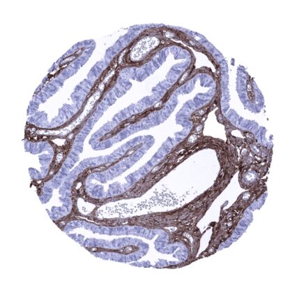
Recombinant Rabbit monoclonal / Rabbit IgG 1:100 – 1:200 Research Use Only Secreted Human MSVA-537R Bone proteoglycan II; CSCD; Dermatan sulphate proteoglycans II (DSPG2); PG40; PGII; PGS2; Proteoglycan core protein; SLRR1B; Small leucine rich protein 1B Fallopian tube: A strong staining should be seen in the stroma while epithelial cells remain negative. Fallopian tube: Epithelial cells should not show any staining. Decorin is an extracellular matrix protein with multiple functions. Decorin is a proteoglycan that weights 90 – 140 kDa and which is coded by the DCN gene on chromosome 12q21.33. Decorin is a member of the small leucine-rich proteoglycan (SLRP) family and represents a significant molecule of the extracellular matrix. It is mainly expressed by f ibroblasts and myofibroblasts. Decorin is closely related to biglycan, another component of connective tissue involved in matrix assembly. Decorin appears to influence fibrillogenesis but also interacts with multiple signaling proteins such as fibronectin, thrombospondin, the complement component C1q, and transforming growth factor-beta (TGF-beta) as well as with various receptor tyrosine kinases such as epidermal growth factor receptor (EGFR), Met, and VEGFR. Through these interactions decorin exerts a multitude of onco suppressive functions including growth inhibition, tumor cell mitophagy, and angiostasis. Decorin also has a pro-inflammatory role through modulation of macrophage function and cytokine secretio... Decorin immunostaining is seen in the stroma of most normal tissues although its quantity is variable. Particularly high levels of staining can be seen along smooth muscle cells, in the myometrium, the stroma of the placenta, endometrium, and the ovary. An intracellular decorin immunostaining occurs in decidua cells (strong, in all cells) and can also be seen in heart muscle and – rarely – in epithelial cells, potentially in case of a focal tissue damage. Squamous epithelial cells – mainly in the granular layer – can show a weak to moderate cytoplasmic staining. The findings described above are this consistent with the RNA data described in the Human Protein Atlas (Tissue expression Decorin) Positive control = Fallopian tube: A strong staining should be seen in the stroma while epithelial cells remain negative. Negative control = Fallopian tube: Epithelial cells should not show any staining. Placenta, mature, amnion and chorion – Strong decorin staining of stroma and possibly decidua c... A decorin immunostaining of variable intensity can be seen in the stroma of cancers from various different entities. More rarely, decorin expression can also occur in the cytoplasm of tumor cells. It has been suggested that a low level of decorin staining in the tumor stroma may be linked to patient prognosis. The TCGA findings on Decorin RNA expression in different tumor categories have been summarized in the Human Protein Atlas. Adenocarcinoma with intense decorin immunostaining of the stroma. A subset of tumor cells – mostly adjacent to the stroma – are also strongly positive. Decorin negative muscle-invasive urothelial carcinoma with strong decorin staining of the stroma. Gastric adenocarcinoma (intestinal type) with moderate to strong decorin staining of tumor cells. Weak decorin positivity of the stroma. Cancer tissue gallery No data available at the moment IHC users have different preferences on how the stains should look like. Some prefer high staining intensity of the target stain and even accept some background. Others favor absolute specificity and lighter target stains. Factors that invariably lead to more intense staining include higher concentration of the antibody and visualization tools, longer incubation time, higher temperature during incubation, higher temperature and longer duration of the heat induced epitope retrieval (slide pretreatment). The impact of the pH during slide pretreatment has variable effects and depends on the antibody and the target protein. All images and data shown here and in our image galleries are obtained by the manual protocol described below. Other protocols resulting in equivalent staining are described as well. Manual protocol Freshly cut sections should be used (less than 10 days between cutting and staining). Heat-induced antigen retrieval for 5 minutes in an autoclave at 121°C in pH 7,8 Target ... The clinical significance of decorin interactions is not clarified yet. The prognostic impact of decorin immunostaining of cancer cells and tumor stroma is unclear. There are two ways how the specificity of antibodies can be documented for immunohistochemistry on formalin fixed tissues. These are: 1. Comparison with a second independent method for target expression measurement across a large number of different tissue types (orthogonal strategy), and 2. Comparison with one or several independent antibodies for the same target and showing that all positive staining results are also seen with other antibodies for the same target (independent antibody strategy). Orthogonal validation: Given the abundant expression of decorin in various tissues, the protein is not well suited for orthogonal validation. The comparison of the MSVA-537R immunostaining data with expression data from three independent RNA screening studies, including the Human Protein Atlas (HPA) RNA-seq tissue dataset, the FANTOM5 project, and the Genotype-Tissue Expression (GTEx) project, which are all summarized in the Human Protein Atlas (Tissue expression Decorin) revealed a good conc...