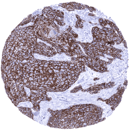
Desmoglein 3 (MSVA-543M)
Mouse monoclonal / IgG 1:75 – 1:150 Research Use Only Cell Surface. Human MSVA-543M 130kDa pemphigus vulgaris antigen (PVA); Balding (Bal); Cadherin family member 6 (CDHF6); Desmoglein-3 (DSG3) Tonsil: A moderate to strong desmoglein 3 immunostaining should be seen in a fraction of squamous epithelial cells. Tonsil: Desmoglein 3 immunostaining should be absent in all non-squamous epithelial cell types. Desmoglein 3 is expressed in squamous epithelium and cells undergoing squamous differentiation. Desmoglein-3 (Dsg3) is a 130 kDa calcium-binding transmembrane glycoprotein which is coded by the DSG3 gene at chromosome 18q12.1. Dsg3, a cadherin superfamily member, is an adhesion protein in desmosomes. In addition to its significant role in cell-cell adhesion, Dsg3 acts as a regulator of pathways governing proliferation, differentiation, and migration of keratinocytes. Because of its suppression of the function of p53, Dsg3 is considered a keratinocyte anti-stress protein. Dsg3 is down-regulated in pemphigus vulgaris where Dsg3 serves as a major antigen (PVA) for autoantibodies, causing disruption of cell–cell cohesion and pemphigus acantholysis in Dsg3-expressing tissues. Dsg3 has also been suggested to play a role in the progression and metastasis of cancer where it was found upregulated. Desmoglein 3 staining pattern in Normal Tissues with antibody MSVA-543M (images are shown in our “Normal Tissue Gallery”) Brain Cerebrum Negative. Cerebellum Negative. Endocrine Tissues Thyroid Negative. Parathyroid Negative. Adrenal gland Negative. Pituitary gland Negative. Respiratory system Respiratory epithelium Weak to moderate Dsg3 staining of a subset of respiratory epithelial cells. Lung Negative. Gastrointestinal Tract Salivary glands Weak to moderate Dsg3 staining in some basal cell layers of small excretory ducts. Esophagus Moderate to strong membranous Dsg3 staining of squamous epithelial cells. The staining intensity decreases towards the surface of the epithelium. Stomach Negative. Duodenum Weak to moderate apical membranous staining of a subset crypt epithelial cells. Small intestine Weak to moderate apical membranous staining of a subset crypt epithelial cells. Appendix Negative. Colon Negative. Rectum Negative. Liver Negative. Gallbladder Negative. Pancreas Negative. G... A positive desmoglein 3 immunostaining is usually seen in squamous cell carcinomas irrespective of their site of origin and in tumor areas exhibiting squamous differentiation. Dsg3 immunostaining can also be seen in various other tumor entities including urothelial cancer, gastric cancer, pancreatic adenocarcinoma, and endometrioid carcinoma. It for example occurs in areas of squamous differentiation. The TCGA findings on Desmoglein 3 RNA expression in different tumor categories have been summarized in the Human Protein Atlas. Esophageal squamous cell carcinoma with strong Dsg3 positivity of tumor cells. Pulmonary squamous cell carcinoma with strong membranous Dsg3 immunostaining of all tumor cells. Squamous cell carcinoma of the penis with intense membranous Dsg3 immunostaining of tumor cells. Cancer tissue gallery Desmoglein 3 (MSVA-543M) publication summary Relevant publication: Viehweger et al. “Desmoglein 3 (Dsg3) expression in cancer: A tissue microarray study on 15,869 tumors” Published in Pathology – Research and Practice 2022 Dec;240:154200. PMID: 36375372. A total of 14’094 tumors from 137 different tumor categories were successfully analyzed by using the following protocol: Heat-induced antigen retrieval for 5 minutes in an autoclave at 121°C in pH 7,8 Target Retrieval Solution buffer. MSVA-543M at a dilution of 1:150 at 37°C for 60 minutes. Visualization of bound antibody by the EnVision Kit (Dako, Agilent). This protocol was also used for all stainings depicted in our tumor and normal tissue galleries. Overall, 47 (34,3%) of 137 tumor categories showed detectable Dsg3 expression in at least one case and 15 (10,9%) tumor categories included at least one case with strong Dsg3 positivity. The highest rate of Dsg3 staining occurred in squamous cell carcinomas from various sites of origin ... IHC users have different preferences on how the stains should look like. Some prefer high staining intensity of the target stain and even accept some background. Others favor absolute specificity and lighter target stains. Factors that invariably lead to more intense staining include higher concentration of the antibody and visualization tools, longer incubation time, higher temperature during incubation, higher temperature and longer duration of the heat induced epitope retrieval (slide pretreatment). The impact of the pH during slide pretreatment has variable effects and depends on the antibody and the target protein. All images and data shown here and in our image galleries are obtained by the manual protocol described below. Other protocols resulting in equivalent staining are described as well. Manual protocol Freshly cut sections should be used (less than 10 days between cutting and staining). Heat-induced antigen retrieval for 5 minutes in an autoclave at 121°C in pH 7,8 Target ... The diagnostic utility of desmoglein 3 IHC should be investigated in a large cohort of tumors from different entities. Desmoglein 3 has been suggested as a prognostic parameter in several different cancer types. This subject deserves further investigation. There are two ways how the specificity of antibodies can be documented for immunohistochemistry on formalin fixed tissues. These are: 1. comparison with a second independent method for target expression measurement across a large number of different tissue types (orthogonal strategy), and 2. Comparison with one or several independent antibodies for the same target and showing that all positive staining results are also seen with other antibodies for the same target (independent antibody strategy). Orthogonal validation: Data derived from three independent RNA screening studies, including the Human Protein Atlas (HPA) RNA-seq tissue dataset, the FANTOM5 project, and the Genotype-Tissue Expression (GTEx) project, which are all summarized in the Human Protein Atlas (Tissue expression Desmoglein 3) suggested that desmoglein 3 expression only occurs in tissues that are covered by squamous epithelium. This fits very well with protein expression data obtained by MSVA-543M . In addition, a wea...