Estrogen Receptor (MSVA-564R)
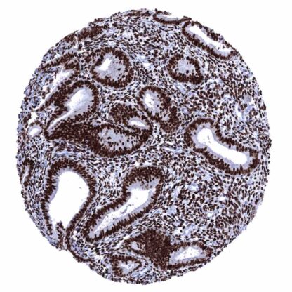
Recombinant Rabbit monoclonal / IgG 1:100-1:200 Research Use Only Nucleus Human MSVA-564R Estrogen Receptor alpha delta 4*5,6,7*/654 isoform;Estrogen Receptor alpha delta 4 +49 isoform; Nuclear receptor subfamily 3 group A member 1 Uterine cervix: virtually all squamous and endocervical cells must show a moderate to strong nuclear staining. Tonsil: Scattered follicular dendritic cells in germinal centers and some squamous epithelial cells in tonsil crypts must show an at least weak nuclear ER immunostaining. Tonsil: B-cells in mantle zones and within germinal centers must be negative. Estrogen receptor is expressed in estrogen dependent cell types. Estrogen receptor alpha (ERα) is a nuclear receptor that is coded by the gene ESR1 located at chromosome 6q25.1. ERα is a ligand-activated transcription factor composed of several domains for hormone binding, DNA binding, and activation of transcription. The classical model of ERα function is that its activation is triggered by estrogen binding to the ERα in the cytoplasm. ERα then dimerizes, translocates to the nucleus, binds to DNA and recruits the necessary transcriptional coregulators that are needed to regulate the transcription of target genes. Evidence suggests that Erα has additional – less well understood – functions that may involve the establishment of chromatin loops and nonnuclear signaling. Irrespective of the exact mechanisms, ERα plays a pivotal role in the development and function of multiple organ systems, including the reproductive (female and male), central nervous, skeletal, and cardiovascular systems. Accordingly, ERα is widely expressed throughout the body. Erα i... The strongest expression of estrogen receptor alpha (ER) is found in the female genital tract including decidual, epithelial and stromal cells of the endometrium and the endocervix, stromal cells and squamous epithelial cells of the ectocervix (lower 2/3), the myometrium, epithelial and stromal cells of the fallopian tube, stromal cells and corpus luteum of the ovary, and in a fraction of the epithelial cells of the breast gland. A moderate to strong ER immunostaining also occurs in stromal cells of the prostate and the seminal vesicles, epithelial cells of the cauda epididymis, peripheral germinative cell of sebaceous glands, peripheral cells of hair follicles, stroma cells of urinary bladder, as well as in a fraction (10-30%) of epithelial cells in the adenohypophysis. A variable, weak to moderate ER positivity can occasionally be seen in a fraction of adrenocortical cells, epithelial cells of salivary glands, smooth muscle cells in the aortic wall, few islet cells and some other cel... The TCGA database on RNA expression in cancer has described the highest levels of ER expression in breast and endometrial cancer followed by ovarian, cervical, and thyroid cancer. Most other important tumor entities are described to be usually “ER negative”. The TCGA findings on Estrogen Receptor RNA expression in different tumor categories have been summarized in the Human Protein Atlas. Endometroid ovarian carcinoma with strong ER immunostaining of tumor cells. Serous high-grade ovarian carcinoma with moderate to strong ER immunostaining of most tumor cells. Invasive breast cancer of no special type (NST) depicting strong ER immunostaining of tumor cells. Cancer tissue gallery Estrogen receptor (MSVA-564R) publication summary Relevant publication: Viehweger et al. “ Estrogen receptor expression in human tumors: A tissue microarray study evaluating more than 18,000 tumors from 149 different entities ” Published in Hum Pathol. 2025 Mar 5;157:105757. doi: 10.1016/j.humpath.2025.105757. Epub ahead of print. PMID: 40054585. A total of 15,960 tumors from 149 different tumor categories were successfully analyzed by using the following protocol: Heat-induced antigen retrieval for 5 minutes in an autoclave at 121°C in pH 9,0 Target Retrieval Solution buffer. MSVA-564R, at a dilution of 1:100 at 37°C for 60 minutes. Visualization of bound antibody by the EnVision Kit (Dako, Agilent). This protocol was also used for all staining’s depicted in our tumor and normal tissue galleries. Overall, 55 of 149 tumor categories showed detectable estrogen receptor (ER) staining with 26 tumor categories showing at least one strongly positive case. ER positivity strongly predominated... IHC users have different preferences on how the stains should look like. Some prefer high staining intensity of the target stain and even accept some background. Others favor absolute specificity and lighter target stains. Factors that invariably lead to more intense staining include higher concentration of the antibody and visualization tools, longer incubation time, higher temperature during incubation, higher temperature and longer duration of the heat induced epitope retrieval (slide pretreatment). The impact of the pH during slide pretreatment has variable effects and depends on the antibody and the target protein. All images and data shown here and in our image galleries are obtained by the manual protocol described below. Other protocols resulting in equivalent staining are described as well. Manual protocol Freshly cut sections should be used (less than 10 days between cutting and staining). Heat-induced antigen retrieval for 5 minutes in an autoclave at 121°C in pH 9 Target Re... ERα is an important drug target in breast cancer. Various new inhibitory molecules are still under development. Because of partly controversial data, the diagnostic utility of ER IHC should be investigated in a large cohort of tumors from different entities. The functional effects of plasma membrane ERα and its role in nonnuclear signaling needs to be further investigated. The molecular basis for tissue-type specific roles of ERα and of tissue-type specific effects of ERα inhibitors is still not fully understood. There are two ways, how the specificity of antibodies can be documented for immunohistochemistry on formalin fixed tissues. These are: There are two ways how the specificity of antibodies can be documented for immunohistochemistry on formalin fixed tissues. These are: 1. comparison with a second independent method for target expression measurement across a large number of different tissue types (orthogonal strategy), and 2. Comparison with one or several independent antibodies for the same target and showing that all positive staining results are also seen with other antibodies for the same target (independent antibody strategy). Orthogonal validation: For the antibody MSVA-564R specificity is suggested by the concordance of the immunostaining data with data from three independent RNA screening studies, including the Human Protein Atlas (HPA) RNA-seq tissue dataset, the FANTOM5 project, and the Genotype-Tissue Expression (GTEx) project, which are all summarized in the Human Protein Atl...