FABP1 (MSVA-501M)
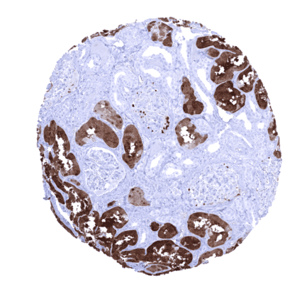
Mouse monoclonal / IgG 1:100-200 Research Use Only Cytoplasmic Human MSVA-501M FABP1, L-FABP Kidney: A strong cytoplasmic FABP1 immunostaining should be seen in cells of the proximal tubule while other cell types remain negative or display only a faint staining. Tonsil: All epithelial and lymphocytic cells must not show any FABP1 immunostaining. FABP1 is expressed in hepatocytes, kidney, and gastrointestinal epithelium. Fatty acid binding protein 1 (FABP1) is a 14kDa protein coded by the FABP1 gene at 2p11.2. FABP1 is found in the liver where it is expressed at high levels, with the expression level reflecting the rate of fatty acid metabolism. There are 9 different isoforms with tissue specific expression in the liver (L-FABP/FABP1), intestinal (I-FABP/FABP2), heart (H-FABP/FABP3), adipocyte (A-FABP/FABP4/aP2), epidermal (E-FABP/FABP5/mal1), ileal (Il-FABP/FABP6), brain (B-FABP/FABP7), myelin (M-FABP/FABP8), and testis (T-FABP/FABP9). Sequence similarity between different FABP isoforms is only 15–70% but all FABPs share a similar three-dimensional structure which enables a binding to various fatty acids. FABP1 is involved in the uptake, intracellular transport and metabolism of long-chain fatty acids, endocannabinoids, phytocannabinoids and other hydrophobic molecules in hepatocytes. In contrast to other fatty acid binding proteins, the FABP1 protein is characterized by a large hydrophobic pocket th... FABP1 staining pattern in Normal Tissues with antibody MSVA-501M (images are shown in our “Normal Tissue Gallery”) Brain Cerebrum Negative. Cerebellum Negative. Endocrine Tissues Thyroid Negative. Parathyroid Negative. Adrenal gland Negative. Pituitary gland Negative. Respiratory system Respiratory epithelium Negative. Lung Negative. Gastrointestinal Tract Salivary glands Negative. Esophagus Negative. Stomach Surface epithelium is usually FABP1 negative, but focal positivity may occur in the antrum. Duodenum Strong FABP1 staining of epithelial cells. The FABP1 staining intensity is strongest at the surface epithelium and decreases towards the crypt bases. Crypt bases may be FABP1 negative. Small intestine Strong FABP1 staining of epithelial cells. The FABP1 staining intensity is strongest at the surface epithelium and decreases towards the crypt bases. Crypt bases may be FABP1 negative. Appendix Strong FABP1 staining of epithelial cells. The FABP1 staining intensity is strongest at the... The TCGA database on RNA expression in cancer has described upregulation of FABP1 in most hepatocellular carcinomas and a fraction of colorectal and stomach cancers. Most other important tumor entities are usually negative. The TCGA findings on FABP1 RNA expression in different tumor categories have been summarized in the Human Protein Atlas. Colorectal adenocarcinoma with very strong FABP1 immunostaining of tumor cells. Contamination (spill over) artifact leads to significant staining of adjacent tumor stroma. Hepatocellular carcinoma with strong FABP1 immunostaining of >95% of tumor cells. Gastric adenocarcinoma (intestinal type) with strong FABP1 immunostaining of most tumor cells. Cancer tissue gallery FABP1 (MSVA-501M) publication summary Relevant publication: Dum et al.: “FABP1 expression in human tumors: A tissue microarray study on 17,071 tumors.” Published in Virchows Archiv Aug 11. doi: 10.1007/s00428-022-03394-5. Epub ahead of print. PMID: 35951102. A total of 14’597 tumors from 150 different tumor categories were successfully analyzed by using the following protocol: Heat-induced antigen retrieval for 5 minutes in an autoclave at 121°C in pH 7,8 Target Retrieval Solution buffer. MSVA-501M at a dilution of 1:150 at 37°C for 60 minutes. Visualization of bound antibody by the EnVision Kit (Dako, Agilent). This protocol was also used for all stainings depicted in our tumor and normal tissue galleries. Overall, 24 (16%) of 150 tumor categories showed detectable FABP1 expression in at least one case and 17 (11%) tumor categories included at least one case with strong FABP1 positivity. By far the highest positivity rates were seen in colorectal adenomas (44-88%), colorectal adenoca... IHC users have different preferences on how the stains should look like. Some prefer high staining intensity of the target stain and even accept some background. Others favor absolute specificity and lighter target stains. Factors that invariably lead to more intense staining include higher concentration of the antibody and visualization tools, longer incubation time, higher temperature during incubation, higher temperature and longer duration of the heat induced epitope retrieval (slide pretreatment). The impact of the pH during slide pretreatment has variable effects and depends on the antibody and the target protein. All images and data shown here and in our image galleries are obtained by the manual protocol described below. Other protocols resulting in equivalent staining are described as well. Manual protocol Freshly cut sections should be used (less than 10 days between cutting and staining). Heat-induced antigen retrieval for 5 minutes in an autoclave at 121°C in pH 7,8 Target ... The diagnostic utility of FABP1 IHC should be investigated in a large cohort of tumors from different entities. Clinical and prognostic significance of FABP1 expression levels in gastrointestinal cancers is unknown. The clinical significance of FABP1 gene variants is under investigation.