Factor XIII alpha (MSVA-813R)
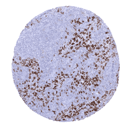
Recombinant Rabbit monoclonal / IgG 1:100 – 1:200 Research Use Only Cytoplasmic and Secreted Human MSVA-813R Coagulation factor XIII A chain; Coagulation factor XIII A1 polypeptide; F13A; F13a1; Fibrin stabilizing factor, A subunit; Fibrinoligase; FSF, A subunit; Protein-glutamine gamma-glutamyltransferase A chain; TGase; Transglutaminase. plasma Colon: A fraction of monocytic cells in the mucosa should show a strong cytoplasmic factor XIIIa immunostaining. Colon:Factor XIIIa immunostaining should be absent in epithelial, endothelial and smooth muscle cells. Factor XIIIa is a part of the blood coagulation system. Factor XIII is a transglutaminase that circulates in the human blood as a heterotetramer of two A and two B subunits. Factor XIIIa is a dimer of activated A peptides. In the process of blood coagulation, factor XIIIa has the function to crosslink fibrin. Factor XIIIs bind to the clot via their B units. In the presence of fibrins, thrombin efficiently cleaves a specific peptide bond of each A unit and activates them. Apart from coagulation and thrombosis, Factor XIII also plays a role in wound healing, bone metabolism, pregnancy and tumor biology. Factor XIIIa is a part of the blood coagulation system. Factor XIII is a transglutaminase that circulates in the human blood as a heterotetramer of two A and two B subunits. Factor XIIIa is a dimer of activated A peptides. In the process of blood coagulation, factor XIIIa has the function to crosslink fibrin. Factor XIIIs bind to the clot via their B units. In the presence of fibrins, thrombin efficiently cleaves a specific peptide bond of each A unit and activates them. Apart from coagulation and thrombosis, Factor XIII also plays a role in wound healing, bone metabolism, pregnancy and tumor biology. Factor XIIIa occurs in monocytes and macrophages of all tissues including dermal dendrocytes and placenta macrophages, megakaryocytes, platelets, chondrocytes, osteoblasts, and osteocytes. These findings are largely consistent with the RNA and protein data described in the Human Protein Atlas (Tissue expression Factor XIIIa) Positive control: Colon: A fraction of monocytic cells in the mucosa should show a strong cytoplasmic factor XIIIa immunostaining. Negative control: Colon: Factor XIIIa immunostaining should be absent in epithelial, endothelial and smooth muscle cells. A strong factor XIIIa immunostaining of mucosa associated macrophages is seen in the colon. Numerous factor XIIIa positive macrophages are seen in the adrenal gland. Macrophages of the first trimenon placenta show a strong factor XIIIa immunostaining. Normal tissue gallery Factor XIIIa is expressed in tumors of monocytic/histiocytic origin. Factor XIIIa immunostaining is often seen in a fraction of tumor associated macrophages. The TCGA findings on Factor XIIIa RNA expression in different tumor categories have been summarized in the Human Protein Atlas. Adrenocortical adenoma containing numerous factor XIIIa positive macrophages. Giant cell tumor of the tendon sheath containing numerous factor XIIIa positive cells. Hodgkin‘s lymphoma containing a dense population of factor XIIIa positive cells. Cancer tissue gallery No data available at the moment IHC users have different preferences on how the stains should look like. Some prefer high staining intensity of the target stain and even accept some background. Others favor absolute specificity and lighter target stains. Factors that invariably lead to more intense staining include higher concentration of the antibody and visualization tools, longer incubation time, higher temperature during incubation, higher temperature and longer duration of the heat induced epitope retrieval (slide pretreatment). The impact of the pH during slide pretreatment has variable effects and depends on the antibody and the target protein. All images and data shown here and in our image galleries are obtained by the manual protocol described below. Other protocols resulting in equivalent staining are described as well. Manual protocol Freshly cut sections should be used (less than 10 days between cutting and staining). Heat-induced antigen retrieval for 5 minutes in an autoclave at 121°C in pH 7,8 Target ... The role of factor XIIIa and of factor XIIIa expressing tumor associated macrophages in cancer biology should be further evaluated. There are two ways how the specificity of antibodies can be documented for immunohistochemistry on formalin fixed tissues. These are: 1. comparison with a second independent method for target expression measurement across a large number of different tissue types (orthogonal strategy), and 2. Comparison with one or several independent antibodies for the same target and showing that all positive staining results are also seen with other antibodies for the same target (independent antibody strategy). Orthogonal validation is not well suited for proteins that are expressed in cells occurring in the majority of all organs such as monocytes and macrophages. RNA expression data derived from the Human Protein Atlas (HPA) RNA-seq tissue dataset, the FANTOM5 project, and the Genotype-Tissue Expression (GTEx) project, which are all summarized in the Human Protein Atlas (Tissue expression Factor XIIIa) are consistent, however, with the staining properties of MSVA-813R . Highest RNA expression leve...