MTAP (MSVA-741R)
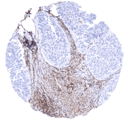
Recombinant Rabbit monoclonal / IgG 1:50 – 1:100 Research Use Only Intracellular Human MSVA-741R BDMF; DMSFH; DMSMFH; Epididymis luminal protein 249; HEL249; LGMBF; MeSAdo phosphorylase; Methylthioadenosine phosphorylase; MSAP; MTA phosphorylase; MTAPase; S- methyl-5”-thioadenosine phosphorylase Ovary: At least a moderate, nuclear and/or cytoplasmic MTAP staining should be seen in ovarian stroma cells. Bladder cancer with homozygous 9p deletion: MTAP staining should be absent in cells from urothelial tumors with homozygous 9p deletion. MTAP is a Sensitive and specific marker for homozygous 9p21 deletions. S-methyl-5′-thioadenosine phosphorylase (MTAP) is an enzyme with a major role in polyamine metabolism. It supports the recovery of both adenine and methionine by catalyzing the phosphorylation of methylthioadenosine (MTA) to adenine and 5-methylthioribose-1-phosphate. Adenine is one of the purine bases required for both DNA and RNA. As an essential protein for the adenine synthesis in human cells, MTAP plays a critical indirect role for the synthesis of DNA and RNA. The MTAP gene is located at 9p21.3. This is in the immediate vicinity of the CDKN2A gene which is homozygously deleted in about 15% of all human cancers. Homozygous co-deletion of MTAP occurs in 80%–90% of tumors with CDKN2A deletion. MTAP deletion have a significant impact on purine biosynthesis because the MTAP substrate MTA accumulates in affected cells and inhibits PRMT5. As a result critical vulnerabilities of affected cells to targeting of the MAT2A/PRMT5/RIOK1 axis develop. Inhibitors of these enzymes are now being t... Images describing the MTAP staining pattern in normal tissues obtained by the antibody MSVA-741R are shown in our “ Normal Tissue Gallery ”. Brain Cerebrum Faint to moderate staining in small blood vessels and in some glia cells. Cerebellum Faint to moderate staining in small blood vessels and in some glia cells. Endocrine Tissues Thyroid Moderate to strong nuclear and cytoplasmic MTAP positivity of epithelial cells. Parathyroid Weak to moderate, predominantly cytoplasmic MTAP positivity of epithelial cells. Adrenal gland Moderate to strong nuclear and cytoplasmic MTAP positivity of epithelial cells. Pituitary gland Weak to strong, predominantly cytoplasmic MTAP positivity of a subset of epithelial cells. Weak, predominantly cytoplasmic MTAP staining of pituicytes of the neurohypophysis. Respiratory system Respiratory epithelium Variable, weak to strong nuclear and cytoplasmic MTAP positivity of respiratory epithelial cells and of bronchial gland cells. Lung Weak to moderate, predomina... Complete loss of MTAP expression occurs in a fraction of cases in urothelial dysplasia, urothelial cancer, malignant mesotheliomas, as well as – less commonly – in various other tumor entities. The TCGA findings on MTAP RNA expression in different tumor categories have been summarized in the Human Protein Atlas. Dysplastic surface urothelium with complete absence of MTAP staining in all urothelial cells. MTAP positive inflammatory cells serve as an internal control Urothelial carcinoma strong nuclear and cytoplasmic MTAP staining of all tumor cells Urothelial carcinoma with complete absence of MTAP staining in all tumor cells. MTAP positive stroma cells serve as an internal control Cancer tissue gallery MTAP (MSVA-741R) publication summary Relevant publication: Gorbokon et al. “Prevalence of S-methyl-5′-thioadenosine Phosphorylase (MTAP) Deficiency in Human Cancer: A Tissue Microarray Study on 13,067 Tumors From 149 Different Tumor Types” Published in Am J Surg Pathol. 2024 Aug 12. Epub ahead of print. PMID: 39132873. A total of 13’067 tumors from 149 different tumor categories were successfully analyzed in a Dako Omnis automated stainer (Agilent Technologies) using the EnVision FLEX, high pH Kit (Agilent Technologies, #GV800). Slides were deparaffinized with clearify agent (Agilent Technologies, #GC810) and exposed to heat-induced antigen retrieval for 30 minutes at 97 °C in target retrieval solution, high pH reagent (part of Agilent kit #GV800). Primary antibody specific for MTAP (recombinant rabbit monoclonal, MSVA-741R, MS Validated Antibodies GmbH, #5293-741R) was applied at ambient temperature for 30 minutes at a dilution of 1:50. Visualization of bound antibody by the EnVision ... IHC users have different preferences on how the stains should look like. Some prefer high staining intensity of the target stain and even accept some background. Others favor absolute specificity and lighter target stains. Factors that invariably lead to more intense staining include higher concentration of the antibody and visualization tools, longer incubation time, higher temperature during incubation, higher temperature and longer duration of the heat induced epitope retrieval (slide pretreatment). The impact of the pH during slide pretreatment has variable effects and depends on the antibody and the target protein. All images and data shown here and in our image galleries are obtained by the manual protocol described below. Other protocols resulting in equivalent staining are described as well. Manual protocol Freshly cut sections should be used (less than 10 days between cutting and staining). Heat-induced antigen retrieval for 5 minutes in an autoclave at 121°C in pH 7,8 Target ... The diagnostic utility of MTAP immunohistochemistry in tumors and in preneoplastic disease needs to be investigated. The prognostic relevance of MTAP expression in tumors should be explored. The predictive role of homozygous and heterozygous MTAP deletions for cancer treatment by drugs targeting the MAT2A/PRMT5/RIOK1 axis needs to be evaluated. There are two ways how the specificity of antibodies can be documented for immunohistochemistry on formalin fixed tissues. These are: 1. Comparison with a second independent method for target expression measurement across a large number of different tissue types (orthogonal strategy), and 2. Comparison with one or several independent antibodies for the same target and showing that all positive staining results are also seen with other antibodies for the same target (independent antibody strategy). Orthogonal validation: For the antibody MSVA-741R orthogonal validation is not suited due to the ubiquitous expression of the protein. Ubiquitous MTAP protein expression is consistent, however, with data from three independent RNA screening studies, including the Human Protein Atlas (HPA) RNA-seq tissue dataset, the FANTOM5 project, and the Genotype-Tissue Expression (GTEx) project, which are all summarized in the Human Protein Atlas (Tissue expression MTAP) . Comparison of antibodies: True M...