Myelin Basic Protein / MBP (MSVA-390R)
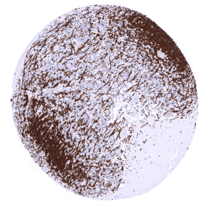
Recombinant Rabbit monoclonal / IgG 1:100 – 1:200 Research Use Only Cell surface and cytoplasm. Human MSVA-390R GDB; Golli MBP; myelin basic protein; Hemopoietic MBP; HMBPR; HUGO; MLD; Myelin A1 Protein, basic; Myelin Deficient; Myelin membrane encephalitogenic protein; SHI; Shiverer; SP Brain: A fibrillar staining pattern should be seen. Colon: MBP staining should be completely absent. MBP is a pivotal protein of myelinated nerves. Myelin basic protein (MBP) is coded by the MBP gene on 18q22. It is produced by oligodendrocytes and plays a relevant role in the myelination of nerves in the nervous system by adhering the cytoplasmic leaflets of the oligodendrocyte (OLG) membrane to each other. MBP maintains the correct structure of myelin, interacts with the lipids in the myelin membrane and forms a ‘molecular sieve’ that restricts diffusion of some membrane proteins from paranodal loops into compact myelin. The gene is characterized by multiple splice variants and a large number of post-translational modifications of the protein, including phosphorylation, methylation, deamidation, citrullination, ADP-ribosylation, and N-terminal acylation. The most common form of MBP is about 18.5 kDa. MBP interacts with various small ligands and proteins. Alterations in these post-translational modifications have been associated with demyelinating diseases. MBP isolated from individuals with multiple sclerosis have had a higher d... MBP is only seen in the brain where MBP immunostaining results in a fibrillar pattern along intracerebral axons. MBP is expressed in oligodendrocytes at a late stage of cell differentiation. These findings are largely consistent with the RNA and protein data described in the Human Protein Atlas (Tissue expression MBP) Positive control: Brain: A fibrillar staining pattern should be seen. Negative control: Colon: MBP staining should be completely absent. Complete absence of MBP immunostaining in epithelial and stromal cells of the appendix mucosa. In the cerebellum a fibrillar MBP immunostaining occurs along intracerebral axons. A dense fibrillar staining is seen in the white matter of the cerebrum. Normal tissue gallery A positive MBP immunostaining is particularly seen in oligodendroglioma and oligoastrocytomas. MBP has been described to be occasionally expressed in teratomas but is otherwise not seen in extracranial tumors. The TCGA findings on MBP RNA expression in different tumor categories have been summarized in the Human Protein Atlas. MBP negative adenocarcinoma of the esophagus. MBP negative prostatic adenocarcinoma (Gleason 3+3=6). MBP negative muscle-invasive plasmacytoid urothelial carcinoma of the urinary bladder. Cancer tissue gallery No data available at the moment IHC users have different preferences on how the stains should look like. Some prefer high staining intensity of the target stain and even accept some background. Others favor absolute specificity and lighter target stains. Factors that invariably lead to more intense staining include higher concentration of the antibody and visualization tools, longer incubation time, higher temperature during incubation, higher temperature and longer duration of the heat induced epitope retrieval (slide pretreatment). The impact of the pH during slide pretreatment has variable effects and depends on the antibody and the target protein. All images and data shown here are obtained by the manual protocol described below. Other protocols resulting in equivalent staining are described as well. Manual protocol Freshly cut sections should be used (less than 10 days between cutting and staining). Heat-induced antigen retrieval for 5 minutes in an autoclave at 121°C in pH 7,8 Target Retrieval Solution buffer. ... MBP is pivotal in research projects that involve an assessment of the status of myelinization of intracranial axons. There are two ways how the specificity of antibodies can be documented for immunohistochemistry on formalin fixed tissues. These are: 1. comparison with a second independent method for target expression measurement across a large number of different tissue types (orthogonal strategy), and 2. Comparison with one or several independent antibodies for the same target and showing that all positive staining results are also seen with other antibodies for the same target (independent antibody strategy). Orthogonal validation: For the antibody MSVA-390R specificity is suggested by the strong concordance of the immunostaining with RNA expression data derived from the Human Protein Atlas (HPA) RNA-seq tissue dataset , the FANTOM5 project, and the Genotype-Tissue Expression (GTEx) project which are all summarized in Human Protein Atlas (Tissue expression MBP) . Immunostaining by using MSVA-390R was almost exclusively detected in the brain, which is the only organ with documented RNA expression. ...