NFIX (HMV329)
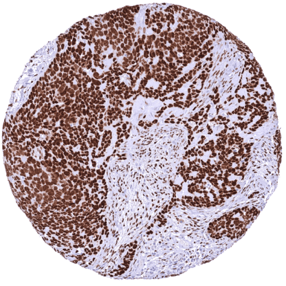
Recombinant Rabbit monoclonal / IgG 1:100 – 1:200 Research Use Only Nucleus Human HMV329 nuclear factor I X , CTF , MRSHSS , NF-I/X , NF1-X , NF1A , SOTOS2 Placenta: A moderate to strong nuclear NFIX staining should be seen in stromal cells while trophoblast cells should remain NFIX negative. Placenta: Trophoblast cells should remain NFIX negative while a moderate to strong nuclear NFIX staining should be seen in stromal cells. NFIX is a master transcription factor interacting with many other transcription factors and chromatin. Nuclear factor 1 X-type (NFIX) is a transcription factor protein which is coded by the NFIX gene on chromosome 19p13.13. Besides NFIA , NFIB, NFIC, NFIX is one out of four closely related members of the nuclear factor I (NFI) family of transcription factors. The family members share a particularly high rate of interactions with other transcription factors. They are therefore thought to not only directly regulate the expression of genes but also believed to modulate the function of other transcription factors. NFIX is known to play a role in muscle and central nervous system embryonic development. For example, NFIX has been shown to control the timing of neural differentiation promoting the ongoing growth of the hippocampus and proper memory function. The role of NFIX in other tissues is less intensively studied. Various types of NFIX alterations have been found in tumors. Mechanisms leading to both increased and reduced expression have been found to promote pro-tumorigenic functions, s... In normal tissues, NFIX is ubiquitously seen in nuclei of all tissues and most cell types. Images describing the NFIX staining pattern in normal tissues obtained by the antibody HMV329 are shown in our “ Normal Tissue Gallery ”. Brain Cerebrum Strong nuclear NFIX staining of glial cells. Weak or absent staining in neurons. Cerebellum Distinct nuclear NFIX staining of glial cells, moderate staining of granular cells, and lack of staining in Purkinje cells. Endocrine Tissues Thyroid Distinct nuclear NFIX staining of follicular cells. Parathyroid Moderate nuclear NFIX staining of all epithelial cells. Adrenal gland Medulla with distinct nuclear NFIX staining of few interspersed stromal (sustentacular cells? endothelial?) cells while adrenal medullary cells remain NFIX negative. Weak to moderate staining of adrenocortical cells. Pituitary gland Distinct nuclear NFIX staining of all pituicytes in the neurohypophysis while only few (epithelial??) cells of the adenohypophysis show a distinct ... A variable level of NFIX staining is seen in many cases of most cancer types. The TCGA findings on NFIX RNA expression in different tumor categories have been summarized in the Human Protein Atlas. Warthin tumor with strong NFIX
positivity of epithelial tumor cells. Laryngeal squamous cell carcinoma
with markedly stronger nuclear
NFIX staining of tumor cells than of
stromal cells. Lymph node containing an NFIX negative
diffuse large B-cell lymphoma. Strong NFIX
staining of interspersed non-neoplastic cells. Cancer tissue gallery No data available at the moment IHC users have different preferences on how the stains should look like. Some prefer high staining intensity of the target stain and even accept some background. Others favor absolute specificity and lighter target stains. Factors that invariably lead to more intense staining include higher concentration of the antibody and visualization tools, longer incubation time, higher temperature during incubation, higher temperature and longer duration of the heat induced epitope retrieval (slide pretreatment). The impact of the pH during slide pretreatment has variable effects and depends on the antibody and the target protein. All images and data shown here and in our image galleries are obtained by the manual protocol described below. Other protocols resulting in equivalent staining are described as well. Manual protocol Freshly cut sections should be used (less than 10 days between cutting and staining). Heat-induced antigen retrieval for 5 minutes in an autoclave at 121°C in pH 7,8 Target ... The role of NFIX in cancer is completely unclear. The patterns of NFIX protein expression in cancer have not been explored. The role of NFIX in heart disease is under investigation. The role of NFIX in other diseases is not specified yet. There are two ways how the specificity of antibodies can be documented for immunohistochemistry on formalin fixed tissues. These are: 1. Comparison with a second independent method for target expression measurement across a large number of different tissue types (orthogonal strategy), and 2. Comparison with one or several independent antibodies for the same target and showing that all positive staining results are also seen with other antibodies for the same target (independent antibody strategy). Orthogonal validation: For the antibody HMV329, specificity is consistent by the consistency of the immunostaining data with data from three independent RNA screening studies, including the Human Protein Atlas (HPA) RNA-seq tissue dataset, the FANTOM5 project, and the Genotype-Tissue Expression (GTEx) project, which are all summarized in the Human Protein Atlas (Tissue expression NFIX) . In agreement with HMV329 immunostaining data, NFIX RNA expression predominated in muscle tissues, was high...