Carbohydrate antigen 19-9 / CA 19-9 (HMV333)
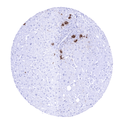
Mouse monoclonal / IgG1 1:100 – 1:200 Research Use Only Intracellular Human HMV333 Liver: A strongCA 19-9staining should be seen in intrahepatic bile ducts (Note that reactivity of control tissues must be previously tested to exclude Le a-b-). Liver:CA 19-9staining should be absent in all hepatocytes. CA 19-9 is a commonly used serum tumor marker. Carbohydrate antigen 19-9 (CA19-9), also termed sialyl-Lewis A, is a cell surface glycoprotein complex which plays a role in cell-to-cell recognition processes. It is a tetrasaccharide carbohydrate containing a transmembrane protein skeleton and extracellular oligosaccharide chains which are extensively glycosylated. CA 19-9 expression is only possible in cells expressing the Lewis gene product, 1,4-fucosyltransferase. As this is only found in patients with Le (a-b+) or Le (a+b-) blood groups there are about 6% of Caucasians and 22% of non-Caucasians who cannot produce CA 19-9. Because CA 19-9 is particularly often overexpressed in pancreatic cancers, serum measurement of CA 19-9 has earlier been recommended for early detection of this cancer. Because of lack of specificity, this approach has largely been abandoned but CA19-9 serum measurement is still used as a marker for disease monitoring. CA19-9 is also being evaluated as a therapeutic target. Images describing the CA 19-9 staining pattern in normal tissues obtained by the antibody HMV333 are shown in our “ Normal Tissue Gallery ”. Brain Cerebrum Negative. Cerebellum Negative. Endocrine Tissues Thyroid Negative. Parathyroid Negative. Adrenal gland Negative. Pituitary gland Negative. Respiratory system Respiratory epithelium In some (but not all samples): Moderate to strong apical membranous CA 19-9 staining of most superficial epithelial cells. In addition, strong membranous and cytoplasmic positivity of a large fraction of goblet cells. Lung Negative. Gastrointestinal Tract Salivary glands Weak to strong, predominantly cytoplasmic (but also membranous) CA 19-9 staining of mucinous glandular cells. Moderate membranous staining of few myoepithelial cells. Weak to moderate membranous CA 19-9 staining of few excretory duct cells. Serous glandular cells are largely negative. Esophagus Strong membranous CA 19-9 staining of a significant fraction of cells in superficial layers of ... CA 19-9 is most expressed in pancreatic, colorectal, gastric, and esophageal adenocarcinomas, mucinous ovarian cancer, cervical adenocarcinoma, cholangiocarcinoma, and urothelial neoplasms, but can also be seen in various other tumor entities. Pancreas – Neuroendocrine tumor with strong CA19-9 positivity of all tumor cells Ovary – CA19-9 negative clear cell carcinoma Uterus, cervix – Adenocarcinoma with intense CA19-9 staining, predominantly seen at the apical pole of tumor cells Cancer tissue gallery No data available at the moment IHC users have different preferences on how the stains should look like. Some prefer high staining intensity of the target stain and even accept some background. Others favor absolute specificity and lighter target stains. Factors that invariably lead to more intense staining include higher concentration of the antibody and visualization tools, longer incubation time, higher temperature during incubation, higher temperature and longer duration of the heat induced epitope retrieval (slide pretreatment). The impact of the pH during slide pretreatment has variable effects and depends on the antibody and the target protein. All images and data shown here and in our image galleries are obtained by the manual protocol described below. Other protocols resulting in equivalent staining are described as well. Manual protocol Freshly cut sections should be used (less than 10 days between cutting and staining). Heat-induced antigen retrieval for 5 minutes in an autoclave at 121°C in pH 7,8 Target ... The diagnostic, prognostic, and predictive role of CA 19-9 expression in tumor tissue and in preneoplastic disease needs to be further clarified. The role of CA 19-9 as a therapeutic target is under investigation. There are two ways how the specificity of antibodies can be documented for immunohistochemistry on formalin fixed tissues. These are: 1. Comparison with a second independent method for target expression measurement across a large number of different tissue types (orthogonal strategy), and 2. Comparison with one or several independent antibodies for the same target and showing that all positive staining results are also seen with other antibodies for the same target (independent antibody strategy). Orthogonal validation: In case of CA 19-9 a comparison with RNA expression cannot be performed because CA 19-9 represents an aberrant glycosylation and not a genuinely expressed gene product. Comparison of antibodies: True presence of CA 19-9 in all cell types for which CA 19-9 staining was observed by HMV333 is corroborated by confirmation of all these CA 19-9 positive cell types by a second independent commercially available anti-CA 19-9 antibody (termed “validation antibody”). HMV333 – App...