CD1a (MSVA-001M)
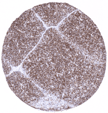
Recombinant Mouse monoclonal / IgG 1:100 – 1:200 Research Use Only Cell surface Human MSVA-001M Cortical thymocyte antigen CD1A, Epidermal dendritic cell marker CD1a antibody, FCB6, HTA1, T cell surface antigen T6 / Leu 6, T-Cell Surface Glycoprotein CD1A Skin: A strong CD1a staining should be seen in Langerhans cells. Skin: CD1a staining must be absent in squamous epithelial cells and sebaceous glands cells. The CD1a protein is a transmembrane glycoprotein encoded by the CD1A gene on 1q23.1. It is a member of a family of 5 CD1 proteins that are structurally related to the major histocompatibility complex (MHC) proteins. It is the main role of CD1 proteins to present lipid and glycolipid antigens of self or microbial origin to T cells. The CD1 family members differ slightly in their specificity for particular lipid ligands in their cellular localization. The CD1a protein is a transmembrane glycoprotein encoded by the CD1A gene on 1q23.1. It is a member of a family of 5 CD1 proteins that are structurally related to the major histocompatibility complex (MHC) proteins. It is the main role of CD1 proteins to present lipid and glycolipid antigens of self or microbial origin to T cells. The CD1 family members differ slightly in their specificity for particular lipid ligands in their cellular localization. CD1a is strongly expressed in Langerhans cells of the skin including hair follicles and sebaceous glands and in non-keratinizing squamous epithelial tissues. A strong CD1a immunostaining is also seen in >80% of the small lymphocytes of the thymic cortex but CD1a is virtually absent in any other lymphatic cells under normal conditions. These findings are largely consistent with RNA and protein data summarized in the Human Protein Atlas (Tissue expression CD1a) . A very faint staining of the surface epithelium of fallopian tube and of endometrium can occasionally be seen and reflects a cross-reactivity. Suggested positive tissue control : S kin: A strong CD1a staining should be seen in Langerhans cells. Suggested negative tissue control: S k in: CD1a staining must be absent in squamous epithelial cells and sebaceous glands cells. Strong CD1a immunostaining in Langerhans cells of the squamous epithelium of the tonsil surface. CD1a immunostaining occurs in >80% of thymocytes in the cortex ... CD1a expression regularly occurs in Langerhans cell histiocytosis but it can also be seen in T cell lymphoma (predominantly cutaneous) and in myeloid leukemia. CD1a positive cells can also be found in regionary lymph nodes where they can migrate. The TCGA findings on CD1a RNA expression in different tumor categories have been summarized in the Human Protein Atlas. Cervival lymph node with a diffuse large B-cell lymphoma showing CD1a positiv dendritic cells. Lymphoblastic T-cell lymphoma showing weak to moderate CD1a immunostaining in a fraction of cells. Hodgkin‘s lymphoma in a supraclavicular lymph node showing scattered CD1a positiv dendritic cells. Cancer tissue gallery No data available at the moment IHC users have different preferences on how the stains should look like. Some prefer high staining intensity of the target stain and even accept some background. Others favor absolute specificity and lighter target stains. Factors that invariably lead to more intense staining include higher concentration of the antibody and visualization tools, longer incubation time, higher temperature during incubation, higher temperature and longer duration of the heat induced epitope retrieval (slide pretreatment). The impact of the pH during slide pretreatment has variable effects and depends on the antibody and the target protein. Accordingly, multiple different protocols can generate identical staining results. All images and data shown here and in our image galleries are obtained by the manual protocol described below. Other protocols resulting in equivalent staining are described as well. Manual protocol Freshly cut sections should be used (less than 10 days between cutting and staining). Heat... The role of CD1a positive cells in inflammatory and allergic disease is not fully understood. The frequency of CD1a immunostaining in Langerhans cell histiocytosis, other hematological and non-hematological neoplasms should be evaluated. Specificity and utility of MSVA-001M is documented by strong positive staining in thymocytes and Langerhans cells, the only cell types known to express CD1a and the absence of CD1a immunostaining in virtually all other types. A very faint staining of the surface epithelium of fallopian tube and of endometrium can occasionally be seen and reflects a cross-reactivity which appears to be negligeable. Normal tissue gallery