SOX10 (MSVA-710R)
Recombinant Rabbit monoclonal / IgG 1:50 – 1:100 Research Use Only Intracellular Human MSVA-710R Transcription factor SOX-10, DOM; PCWH; SOX10; SRY (sex determining region Y) box 10; SRY box containing gene 10; SRY related HMG box gene 10; Waardenburg syndrome type 2E(WS2E); WS4; Waardenburg syndrome type 4C (WS4C) Breast: A moderate to strong nuclear SOX10 staining should be seen in breast epithelial cells. Colon: SOX10 staining must be completely absent in epithelial cells. SOX10 is a epigenetic regulator and therapeutic target protein. SOX10 is one of at least 20 members of the family of SRY (sex-determining region Y)-related high mobility group box-containing (SOX) proteins most of which have a role in embryogenesis and cell differentiation. SOX10 is coded by the SOX10 gene on chromosome 22q13.1. It is critical for the development of neural crest cells, glial cells, Schwann cells, neurons, osteoblasts, smooth muscle cells and melanocytes. SOX10 protein acts as a transcriptional activator after forming a protein complex with other proteins. In melanocytic cells, SOX10 expression is regulated by MITF. Heterozygous germline mutations occurring within and around SOX10 can cause various clinical syndroms characterized by pigment abnormalities, disorders of gastrointestinal motility, loss of smell, and hearing loss. Depending on the type of mutations, Waardenburg syndrome type 4 ( auditory pigmentary abnormalities and Hirschsprung disease), Waardenburg syndrome type 2 (Waardenburg syndrome without Hirschsprung disease), P... SOX10 is a nuclear protein which is preferentially expressed in melanocytes of the skin, glia cells of the cerebrum and the cerebellum, eccrine skin glands, salivary glands, and the breast. A strong nuclear SOX10 staining of scattered spindle shaped stroma cells can be found in various tissues Images describing the SOX10 staining pattern in normal tissues obtained by the antibody MSVA-710R are shown in our “ Normal Tissue Gallery ”. Brain Cerebrum Strong nuclear SOX10 positivity of most glia cells. Cerebellum Strong nuclear SOX10 positivity of most glia cells. Endocrine Tissues Thyroid Negative. Parathyroid Numerous scattered spindle shaped SOX10 positive cells between glandular cells. Adrenal gland Scattered spindle shaped SOX10 positive cells between glandular cells. Pituitary gland Negative. Respiratory system Respiratory epithelium Respiratory epithelium is SOX10 negative. Bronchial glands with a distinct nuclear SOX10 positivity. Scattered spindle shaped SOX10 positive cells in th... SOX10 expression preferentially occurs in gliomas, neoplasms of melanocytes and nerve sheaths, as well as of cancers derived from salivary, breast, and eccrine skin glands. The TCGA findings on SOX10 RNA expression in different tumor categories have been summarized in the Human Protein Atlas. Colorectal adenocarcinoma lacking
SOX10 staining in tumor cells but
containing a small SOX10 positive
nerve. Invasive breast cancer of no special type
(NST) with strong SOX10 staining of all
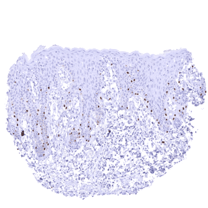
tumor cells. SOX10 negative invasive breast cancer
of no special type (NST). SOX10
staining is seen in few retained normal
breast glands. Cancer tissue gallery No data available at the moment IHC users have different preferences on how the stains should look like. Some prefer high staining intensity of the target stain and even accept some background. Others favor absolute specificity and lighter target stains. Factors that invariably lead to more intense staining include higher concentration of the antibody and visualization tools, longer incubation time, higher temperature during incubation, higher temperature and longer duration of the heat induced epitope retrieval (slide pretreatment). The impact of the pH during slide pretreatment has variable effects and depends on the antibody and the target protein. All images and data shown here and in our image galleries are obtained by the manual protocol described below. Other protocols resulting in equivalent staining are described as well. Manual protocol Freshly cut sections should be used (less than 10 days between cutting and staining). Heat-induced antigen retrieval for 5 minutes in an autoclave at 121°C in pH 7,8 Target ... The prevalence and role of SOX10 expression in different tumor types needs to be further clarified. The function of SOX10 and its role in various diseases needs to be further investigated. The suitability of SOX10 as a therapeutic target needs to be tested. There are two ways how the specificity of antibodies can be documented for immunohistochemistry on formalin fixed tissues. These are: 1. Comparison with a second independent method for target expression measurement across a large number of different tissue types (orthogonal strategy), and 2. Comparison with one or several independent antibodies for the same target and showing that all positive staining results are also seen with other antibodies for the same target (independent antibody strategy). Orthogonal validation: For the antibody MSVA-710R specificity is supported by the good concordance of the immunostaining data with data from three independent RNA screening studies, including the Human Protein Atlas (HPA) RNA-seq tissue dataset, the FANTOM5 project, and the Genotype-Tissue Expression (GTEx) project, which are all summarized in the Human Protein Atlas (Tissue expression SOX10) . SOX10 staining by MSVA-710R was preferably detected in salivary glands, the brain and in breast gla...