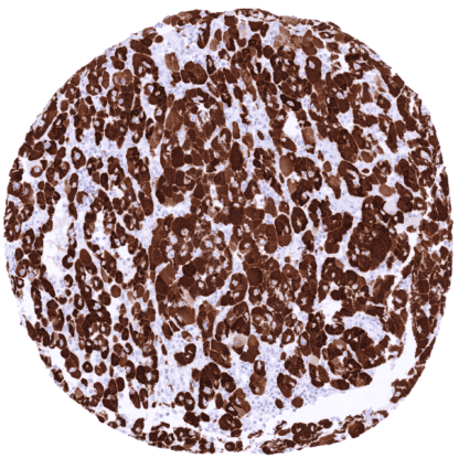
STING (MSVA-515M)
Mouse monoclonal / IgG 1:100 – 1:200 Research Use Only Cytoplasm Human MSVA-515M Endoplasmic reticulum interferon stimulator; ERIS; hMITA; hSTING; Mediator of IRF3 activation; MITA; NET23; MPYS; Mitochondrial mediator of IRF3 activation; N terminal methionine proline tyrosine serine plasma membrane tetraspanner; Stimulator of interferon genes protein; Transmembrane protein 173 Adrenal gland: Endothelial cells and macrophages should show a moderate to strong STING positivity. Adrenal gland: Adrenocortical and medullary cells should not show STING staining (Note: in case of tissue damage aberrant STING staining may occur). STING1 is pivotal protein for innate immunity acting as a pathogen receptor. Stimulator of interferon genes (STING), also known as transmembrane protein 173 (TMEM173) is coded by the STING1 gene on chromosome 5q31 . 2. STING is a pathogen recognition receptor located in the endoplasmic reticulum. It is activated by cyclic dinucleotides (CDNs) which are either derived from microorganisms or synthesized by the enzyme cyclic GMP-AMP synthase (cGAS) in response to binding either host- or pathogen-derived cytosolic double-stranded (ds)DNA. STING plays an important role in innate immunity. STING induces type I interferon production when cells are infected with intracellular pathogens, such as viruses, mycobacteria and intracellular parasites. Type I interferon, mediated by STING, protects infected cells and adjacent cells from infection by autocrine and paracrine signaling. STING works as both a direct cytosolic DNA sensor (CDS) and an adaptor protein in Type I interferon signaling through different molecular mechanisms. STING activates downstream transcription facto... STING staining pattern in Normal Tissues with antibody MSVA-515M (Images shown in our “Normal Tissue Gallery”) Brain Cerebrum STING staining of endothelial cells. Cerebellum STING staining of endothelial cells. Endocrine Tissues Thyroid STING staining of endothelial cells. Parathyroid STING staining of endothelial cells. Adrenal gland STING staining of endothelial cells and of macrophages. Pituitary gland STING staining of endothelial cells and (in some samples) of a subset of epithelial cells. Respiratory system Respiratory epithelium Diffuse strong staining of epithelial cells. Lung Strong STING staining of macrophages. Gastrointestinal Tract Salivary glands STING staining of endothelial cells and of basal cells in excretory ducts. Esophagus Weak to moderate STING staining of the basal cell layers of the squamous epithelium. Stomach Variable STING staining of epithelial cells ranging from complete lack of staining to substantial staining of most or all cells. Duodenum Variable STING ... A variable STING staining of tumor cells and of stroma cells can occur in many different tumor entities. The TCGA findings on STING1 RNA expression in different tumor categories have been summarized in the Human Protein Atlas. Colorectal adenocarcinoma with strong STING immunostaining of tumor cells. Tumor-adjacent inflammatory cells are also strongly positive STING negative colorectal adenocarcinoma. Inflammatory cells and capillaries are STING positive Colorectal adenocarcinoma with variable, weak to moderate STING staining of tumor cells. Inflammatory cells and capillaries show stronger STING staining Cancer tissue gallery STING (MSVA-515M) publication summary: Relevant publication: Menz et al.: “ Stimulator of Interferon Genes Protein (STING) Expression in Cancer Cells: A Tissue Microarray Study Evaluating More than 18,000 Tumors from 139 Different Tumor Entities” Published in Cancers (Basel) 2024, Jun 30 PMID: 39001487 A total of 15’345 tumors were successfully analyzed from 139 different tumor categories by using the following protocol: Heat-induced antigen retrieval for 5 minutes in an autoclave at 121°C in pH7,8 Target Retrieval Solution buffer. Apply MSVA-515M at a dilution of 1:150 at 37°C for 60 minutes. Visualization of bound antibody by the EnVision Kit (Dako, Agilent). This protocol was also used for all stainings depicted in our tumor and normal tissue galleries. Almost all previous studies on STING expression in cancer had only considered STING expression in tumor infiltrating inflammatory cells. The study of Menz et al. therefore entirely focused on STING in tumor cells. At least one STING ... IHC users have different preferences on how the stains should look like. Some prefer high staining intensity of the target stain and even accept some background. Others favor absolute specificity and lighter target stains. Factors that invariably lead to more intense staining include higher concentration of the antibody and visualization tools, longer incubation time, higher temperature during incubation, higher temperature and longer duration of the heat induced epitope retrieval (slide pretreatment). The impact of the pH during slide pretreatment has variable effects and depends on the antibody and the target protein. All images and data shown here and in our image galleries are obtained by the manual protocol described below. Other protocols resulting in equivalent staining are described as well. Manual protocol Freshly cut sections should be used (less than 10 days between cutting and staining). Heat-induced antigen retrieval for 5 minutes in an autoclave at 121°C in pH 7,8 Target ... The diagnostic utility of STING expression analysis is unclear. The prognostic and predictive role of different levels of STING expression in cancer should be investigated. A comparison of the expression of STING across different tumor entities would be of interest. The function of STING is still not completely clear. The utility of STING as a therapeutic target should be evaluated. There are two ways how the specificity of antibodies can be documented for immunohistochemistry on formalin fixed tissues. These are: 1. Comparison with a second independent method for target expression measurement across a large number of different tissue types (orthogonal strategy), and 2. Comparison with one or several independent antibodies for the same target and showing that all positive staining results are also seen with other antibodies for the same target (independent antibody strategy). Orthogonal validation: Given the ubiquitous presence of STING RNA across all different normal tissues and the restriction of STING expression to specific cell types, orthogonal validation is not applicable for STING. Comparison of antibodies: Specificity of MSVA-515M binding for STING is documented by the confirmation of all staining results obtained by MSVA-515M in a parallel analysis using an independent second commercial STING antibody (termed “validation antibody”). MSVA-515M – Adenohypop...