Tyrosine Hydroxylase / TH (HMV312)
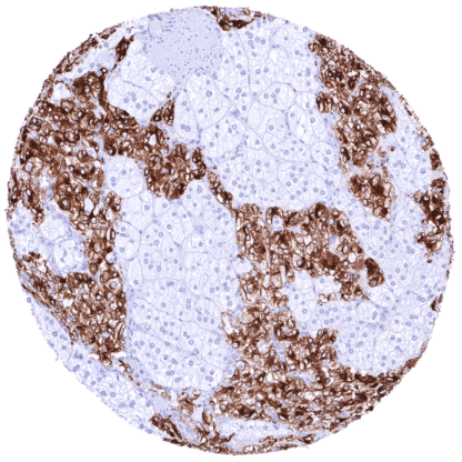
Recombinant Rabbit monoclonal / IgG 1:100 – 1:200 Research Use Only Intracellular Human HMV312 tyrosine hydroxylase , DYT14 , DYT5b , TYH Adrenal gland: A strong cytoplasmic and membranousTHstaining should be seen in medullary cells as well as in a subset of nerve fibers in the cortex. Adrenal gland:TH staining must be absent in adrenocortical cells. Tyrosine Hydroxylase is a Rate limiting enzyme for catecholin synthesis. The enzyme tyrosine hydroxylase (TH) or tyrosine 3-monooxygenase is coded by the TH gene at 11p15.5. TH catalyzes the rate limiting step in the synthesis of catecholamines which is the conversion of L-tyrosine to L-3,4-dihydroxyphenylalanine (L-DOPA). L-DOPA is a precursor for dopamine, which, in turn, is a precursor for the important neurotransmitters norepinephrine (noradrenaline) and epinephrine (adrenaline). These catecholamines play important roles in a variety of physiological and behavioral functions in the nervous and endocrine systems. Tyrosine hydroxylase deficiency leads to impaired synthesis of catecholamines and causes a progressive encephalopathy with poor prognosis. TH is also involved in other neurological diseases. For example, cerebral TH activity is significantly reduced in patients with Alzheimer’s disease as compared to healthy individuals. Tyrosine hydroxylase is also an autoantigen in Autoimmune Polyendocrine Syndrome (APS) type I. In normal tissues, TH is present in specific regions of the central nervous system (CNS), peripheral sympathetic neurons and nerve fibers, as well as in the adrenal medulla. TH is mainly present in the cytosol, although it also is found in the plasma membrane probably due to catecholamine packing in vesicles and export through the synaptic membrane. Images describing the Tyrosine Hydroxylase (TH) staining pattern in normal tissues obtained by the antibody HMV 312 are shown in our “ Normal Tissue Gallery ”. Brain Cerebrum Negative. Cerebellum Negative. Endocrine Tissues Thyroid Negative. Parathyroid Negative. Adrenal gland Strong membranous and cytoplasmic TH staining of medullary cells. Some nerve fibers in the cortex also show distinct TH positivity. Pituitary gland Negative. Respiratory system Respiratory epithelium Negative. Lung Negative. Gastrointestinal Tract Salivary glands Negative. Some nerve fibers exhibit distinct TH positivity. Esophagus Negative. Stomach Negative. Duodenum... TH expression occurs in pheochromocytomas (almost always positive) and paraganglioma (about 30% positive) and rarely in other tumors. The TCGA findings on TH RNA expression in different tumor categories have been summarized in the Human Protein Atlas. Paraganglioma with strong TH staining of tumor cells. Medullary carcinoma of the thyroid showing a small
number of dispersed cells with strong TH positivity. Paraganglioma with strong TH staining of tumor cells. Cancer tissue gallery No data available at the moment IHC users have different preferences on how the stains should look like. Some prefer high staining intensity of the target stain and even accept some background. Others favor absolute specificity and lighter target stains. Factors that invariably lead to more intense staining include higher concentration of the antibody and visualization tools, longer incubation time, higher temperature during incubation, higher temperature and longer duration of the heat induced epitope retrieval (slide pretreatment). The impact of the pH during slide pretreatment has variable effects and depends on the antibody and the target protein. All images and data shown here and in our image galleries are obtained by the manual protocol described below. Other protocols resulting in equivalent staining are described as well. Manual protocol Freshly cut sections should be used (less than 10 days between cutting and staining). Heat-induced antigen retrieval for 5 minutes in an autoclave at 121°C in pH 7,8 Target ... Quantification of TH in brain and nerve tissues. The clinical significance of the level of TH expression in paragangliomas needs to be further evaluated. The specificity of TH immunostaining for pheochromocytoma and paraganglioma needs to be determined. There are two ways how the specificity of antibodies can be documented for immunohistochemistry on formalin fixed tissues. These are: 1. Comparison with a second independent method for target expression measurement across a large number of different tissue types (orthogonal strategy), and 2. Comparison with one or several independent antibodies for the same target and showing that all positive staining results are also seen with other antibodies for the same target (independent antibody strategy). Orthogonal validation: For the antibody HMV312 specificity is suggested by the strong concordance of the immunostaining data with data from three independent RNA screening studies, including the Human Protein Atlas (HPA) RNA-seq tissue dataset, the FANTOM5 project, and the Genotype-Tissue Expression (GTEx) project, which are all summarized in the Human Protein Atlas (Tissue expression TH) . TH positivity by HMV312 is very strong in the only extracranial tissues with documented TH RNA expressi...