Uroplakin 1B (MSVA-734M)
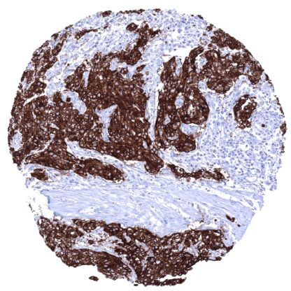
Mouse monoclonal / IgG 1:100 – 1:200 Research Use Only Cell Surface and cytoplasm Human MSVA-734M Tetraspanin-20; Tspan-20; TSPAN20; UP1b; UPIb; UPK1B; Uroplakin-1b Urinary bladder:A strong membranous and cytoplasmic UPK1b immunostaining should be seen in the urothelium (the staining can be limited to the top cell layers or equally involve all cell layers). Colon:UPK1b immunostaining should be absent in all cells of the colon mucosa. Uroplakin 1B is a marker for urothelial carcinomas Uroplakin 1B (Upk1b) is a 29.6 kDa protein which is encoded by the UPK1b gene located at 3q13.3-q21. In humans, there are 4 other uroplakins (Upk 1a, -2, -3a, -3b), which assemble into dimers of Upk1a/Upk2 and Upk1b/Upk3 and eventually into heterotetramers that are deposited at the surface of the epithelial cells lining the urinary bladder, ureter, and renal pelvis. These membrane plaques are termed asymmetric unit membranes (AUMs) and serve as elastic stabilizers that prevent the bladder wall from mechanical stress and rupture during urinary bladder distension. In addition, AUMs may contribute to the regulation of membrane permeability and signal transduction, which may also impact aspects of tumorigenesis such as cellular growth and motility. Uroplakin 1B staining pattern in Normal Tissues with antibody MSVA-734M (Images shown in our “Normal Tissue Gallery”) Brain Cerebrum Negative. Cerebellum Negative. Endocrine Tissues Thyroid Negative. Parathyroid Negative. Adrenal gland Negative. Pituitary gland Negative. Respiratory system Respiratory epithelium A moderate to strong Upk1b staining occurs in a fraction of cells. Lung Gastrointestinal Tract Salivary glands A variable (weak to strong) Upk1b positivity can sometimes occur in some serous cells in salivary glands. Esophagus Usually negative. Scattered Upk1b positive cells can occur. Stomach A moderate to strong Upk1b staining is seen in superficial epithelial cells and parietal cells. Colon Negative. Duodenum Negative. Rectum Negative. Small intestine Negative. Liver A variable (weak to strong) cytoplasmic and membranous Upk1b positivity can sometimes occur in intrahepatic bile ducts. Hepatocytes are negative. Gallbladder A variable (weak to strong) cytoplasmic and membranou... The TCGA database on RNA expression in cancer has described upregulation of Upk1b in a fraction of urothelial, head and neck, lung, endometrial, cervical, ovarian and renal cancers. The TCGA findings on Uroplakin 1B RNA expression in different tumor categories have been summarized in the Human Protein Atlas. Non-invasive urothelial carcinoma (low grade, pTaG2) with strong Upk1b immunostaining of tumor cells. Warthin tumor with strong Upk1b immunostaining of a fraction of tumor cells. Malignant mesothelioma (epitheloid) with strong Upk1b immunostaining of all tumor cells. Cancer tissue gallery Uroplakin 1b (MSVA-734M) publication summary: Relevant publication: Reiswich et al. “ Large-scale human tissue analysis identifies Uroplakin 1b as a putative diagnostic marker in surgical pathology “ Published in Human Pathology 2022 May 9; S0046-8177(22)00116-2. doi: 10.1016/j.humpath.2022.05.002. PMID: 35550834 In this study, 11’868 tumors were analyzed from 127 different tumor categories by using the following protocol: Heat-induced antigen retrieval for 5 minutes in an autoclave at 121°C in pH 7.8 Target Retrieval Solution buffer. MSVA-734M at a dilution of 1:150 at 37°C for 60 minutes. Visualization of bound antibody by the EnVision Kit (Dako, Agilent). This protocol was also used for all stainings depicted in our tumor and normal tissue galleries. At least one case with a positive Upk1b immunostaining was seen in 61 (48%) and at least one case with a strong Upk1b immunostaining was seen in 39 (31%) of 127 tumor categories. The distribution of positive staining results is shown i... IHC users have different preferences on how the stains should look like. Some prefer high staining intensity of the target stain and even accept some background. Others favor absolute specificity and lighter target stains. Factors that invariably lead to more intense staining include higher concentration of the antibody and visualization tools, longer incubation time, higher temperature during incubation, higher temperature and longer duration of the heat induced epitope retrieval (slide pretreatment). The impact of the pH during slide pretreatment has variable effects and depends on the antibody and the target protein. All images and data shown here and in our image galleries are obtained by the manual protocol described below. Other protocols resulting in equivalent staining are described as well. Manual protocol Freshly cut sections should be used (less than 10 days between cutting and staining). Heat-induced antigen retrieval for 5 minutes in an autoclave at 121°C in pH 7,8 Target ... The prevalence and clinical significance of Upk1b expression in cancer is unknown. There are two ways how the specificity of antibodies can be documented for immunohistochemistry on formalin fixed tissues. These are: 1. comparison with a second independent method for target expression measurement across a large number of different tissue types (orthogonal strategy), and 2. Comparison with one or several independent antibodies for the same target and showing that all positive staining results are also seen with other antibodies for the same target (independent antibody strategy). Orthogonal validation: Specificity of the antibody MSVA-734M for Upk1b is suggested by the strong concordance of its immunostaining pattern with data from three independent RNA screening studies, including the Human Protein Atlas (HPA) RNA-seq tissue dataset, the FANTOM5 project, and the Genotype-Tissue Expression (GTEx) project, which are all summarized in the Human Protein Atlas (Tissue expression Uroplakin 1B) . Immunostaining by MSVA-734M is detected in all organs with documented Upk1b RN...