Uroplakin 3B (MSVA-736M)
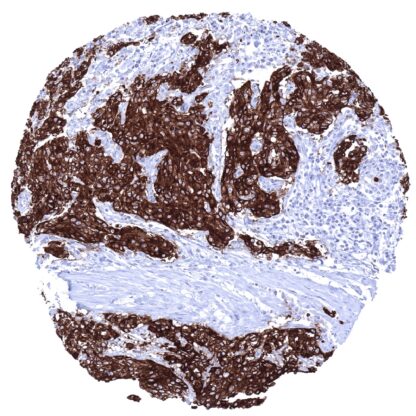
Mouse monoclonal / IgG 1:100 – 1:200 Research Use Only Cell Surface and cytoplasm Human MSVA-736M FLJ32198, MGC10902, p35, UPIIIb Urinary bladder: A moderate to strong membranous Upk3b immunostaining should be seen in umbrella cells of the normal urothelium (the staining is usually limited to apical surface membrane of umbrella cells). Colon: Upk3b immunostaining should be absent in all cells of the colon mucosa. Uroplakin 3B is a marker for mesothelial cells and umbrella cells. The Uroplakin 3B (Upk3b) protein is coded by the UPK3b gene located at 7q1.23 . Upk3b is one out of 5 known uroplakin (Upk) protein particles that cooperatively form apical asymmetrical unit membrane (AUM) plaques which play an important role in the stabilization and strengthening of epithelial cells that line the bladder. These AUM plaques enable the inner bladder membrane to stretch and prevent urothelial cells from rupturing during bladder distension. Upks are assembled in the endoplasmic reticulum (ER), where they heterodimerize prior to escaping the ER. Upk3b heterodimerizes with Upk1b. Upk heterodimers subsequently form tetramers which then combine as concentric hexameric rings that are packaged into vesicles and trafficked to the cell surface. AUMs and Upk proteins may have a role in mediating membrane permeability and signal transduction events that are involved in the regulation of cell development, activation, growth, and motility. Uroplakin 3B staining pattern in Normal Tissues with antibody MSVA-736M (Images shown in our “Normal Tissue Gallery”) Brain Cerebrum Negative. Cerebellum Negative. Endocrine Tissues Thyroid Negative. Parathyroid Negative. Adrenal gland Negative. Pituitary gland Negative. Respiratory system Respiratory epithelium Negative. Lung Negative. Gastrointestinal Tract Salivary glands Negative. Esophagus Negative. Stomach Negative. Duodenum Negative. Small intestine Negative. Appendix Negative. Colon Negative. Rectum Negative. Liver Negative. Gallbladder Negative. Pancreas Negative. Genitourinary Kidney Negative. Urothelium Moderate to strong, membranous Upk3b staining of the apical membrane of umbrella cells. Male genital Prostate Negative. Seminal vesicles Negative. Testis Negative. Epididymis Negative. Female genital Breast Negative. Uterus, myometrium Negative. Uterus, ectocervix Negative. Uterus endocervix Negative. Uterus, endometrium Negative. Fallopian Tube Negative. Ovary Negative. Plac... According to the TCGA data, Upk3b expression is most commonly seen in urothelial carcinomas and in ovarian cancers. Rarely, it can also be found in other tumors. The TCGA findings on Uroplakin 3B RNA expression in different tumor categories have been summarized in the Human Protein Atlas. Muscle-invasive urothelial carcinoma with strong, predominantly membranous Upk3b immunostaining of most tumor cells Serous high-grade carcinoma with strong membranous Upk3b staining in 80% of tumor cells Mesothelioma showing moderate to strong membranous, predominantly apical Upk3b positivity of epitheloid tumor cells Cancer tissue gallery Uroplakin 3b (Upk3b) (MSVA-736M) publication summary Relevant publication: Lennartz et al. : “Analysis of More than 16,000 Human Tumor and Normal Tissues Identifies Uroplakin 3B as a Useful Diagnostic Marker for Mesothelioma and Normal Mesothelial Cells.” Published in Diagnostics (Basel). 2022 Oct 17;12(10):2516. PMID: 36292206 A total of 16’185 tumors were successfully analyzed from 151 different tumor categories by using the following protocol: Heat-induced antigen retrieval for 5 minutes in an autoclave at 121°C in pH7,8 Target Retrieval Solution buffer. MSVA-736M at a dilution of 1:150 at 37°C for 60 minutes. Visualization of bound antibody by the EnVision Kit (Dako, Agilent). This protocol was also used for all stainings depicted in our tumor and normal tissue galleries. In this study, at least one Upk3b positive case was seen 17 of 151 (11.3%) of tumor types and only 6 (4%) tumor categories included at least one case with strong positivity. Lennartz et al described the highest Up... IHC users have different preferences on how the stains should look like. Some prefer high staining intensity of the target stain and even accept some background. Others favor absolute specificity and lighter target stains. Factors that invariably lead to more intense staining include higher concentration of the antibody and visualization tools, longer incubation time, higher temperature during incubation, higher temperature and longer duration of the heat induced epitope retrieval (slide pretreatment). The impact of the pH during slide pretreatment has variable effects and depends on the antibody and the target protein. All images and data shown here and in our image galleries are obtained by the manual protocol described below. Other protocols resulting in equivalent staining are described as well. Manual protocol Freshly cut sections should be used (less than 10 days between cutting and staining). Heat-induced antigen retrieval for 5 minutes in an autoclave at 121°C in pH 7,8 Target ... The prevalence and clinical significance of Upk3b expression in cancer is unknown. The potential diagnostic utility of Upk3b immunostaining needs to be further evaluated. There are two ways how the specificity of antibodies can be documented for immunohistochemistry on formalin fixed tissues. These are: 1. Comparison with a second independent method for target expression measurement across a large number of different tissue types (orthogonal strategy), and 2. Comparison with one or several independent antibodies for the same target and showing that all positive staining results are also seen with other antibodies for the same target (independent antibody strategy). Orthogonal validation: For the antibody MSVA-736M considerable discrepancy exist as compared to data from three independent RNA screening studies, including the Human Protein Atlas (HPA) RNA-seq tissue dataset, the FANTOM5 project, and the Genotype-Tissue Expression (GTEx) project, which are all summarized in the Human Protein Atlas (Tissue expression Uroplakin 3B) . In concordance with MSVA-736M IHC data, RNA expression is seen in the urinary bladder, but other tissues with high RNA expressi...