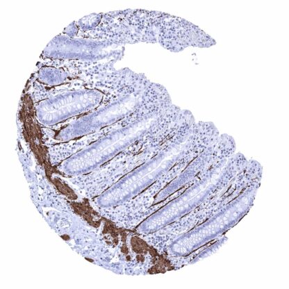
Caldesmon (MSVA-538R)
Recombinant Rabbit monoclonal / Rabbit IgG 1:100 – 1:200 Research Use Only Cytoplasmic Human MSVA-538R CAD; CALD1; Caldesmon 1 Isoform 1; Caldesmon 1 Isoform 2; Caldesmon 1 Isoform 3; Caldesmon 1 Isoform 4; Caldesmon 1 Isoform 5; CDM; HCAD; LCAD; NAG22 Colon: A strong caldesmon staining should be seen of the muscularis mucosa and of a membrane-like layer along the basement membranes of crypts (intestinal subepithelial myofibroblasts). Colon: Normal epithelial cells must not show caldesmon staining. Caldesmon is a sensitive and specific marker for smooth muscle cells. Caldesmon is a particularly long (75 nm) and flexible molecule that is coded by the CALD1 gene located at chromosome 7q33. Together with tropomyosin, it regulates the binding of myosin to actin. In relaxed smooth muscle the actin binding site on myosin is blocked by the caldesmon-tropomyosin complex. Stimulation of smooth muscle results in ERK-dependent phosphorylation of caldesmon and subsequent removal of the caldesmon-tropomyosin complex from the myosin binding site on actin and consequently in muscle contraction. Two alternatively spliced isoforms of caldesmon occur. The heavy isoform (h-Caldesmon) is largely restricted to differentiated contractile smooth muscle cells but the light isoform (L-Caldesmon) also exists in other cell types. The antibody MSVA-538R recognizes h-caldesmon. Caldesmon-h staining is seen in the cytoplasm and cell membrane of smooth muscles in various organs including the walls of small, medium sized, and large vessels, the aortic media and smooth muscle elements of the ovarian stroma. Myoepithelial cells and basal cells of excretory ducts in salivary glands as well as myoepithelial cells of the breast are also caldesmon-h positive. In the colorectum, caldesmon-h stains intestinal subepithelial myofibroblasts (ISEMFs) which form a thin membrane-like layer around the colon crypts. A weak but distinct membranous caldesmon-h staining also occurs at the basal membrane of amnion cells. The findings described above are this consistent with the RNA data described in the Human Protein Atlas (Tissue expression Caldesmon) Positive control = Colon: A strong caldesmon staining should be seen of the muscularis mucosa and of a membrane-like layer along the basement membranes of crypts (intestinal subepithelial myofibroblasts). Negative control = Colon: No... A positive caldesmon immunostaining can be seen in the vast majority of leiomyomas and leiomyosarcomas as well as in other more rare smooth muscle tumors such as myopericytomas or Glomus tumors. The TCGA findings on Caldesmon RNA expression in different tumor categories have been summarized in the Human Protein Atlas. Of note, these data are likely to not reflect h-caldesmon expression of tumor cells. They may rather be driven by the quantity of non-neoplastic smooth muscle invaded by the tumor cells and possibly also include L-caldesmon RNA. Leiomyosarcoma with strong Caldesmon immunostaining of tumor cells. Caldesmon negative adenocarcinoma (Gleason 3+3=6) surrounded by prostatic stroma rich in caldesmon positive smooth muscle cells. Angiomyolipoma with strong caldesmon immunostaining of muscular tumor cells. Cancer tissue gallery No data available at the moment IHC users have different preferences on how the stains should look like. Some prefer high staining intensity of the target stain and even accept some background. Others favor absolute specificity and lighter target stains. Factors that invariably lead to more intense staining include higher concentration of the antibody and visualization tools, longer incubation time, higher temperature during incubation, higher temperature and longer duration of the heat induced epitope retrieval (slide pretreatment). The impact of the pH during slide pretreatment has variable effects and depends on the antibody and the target protein. All images and data shown here and in our image galleries are obtained by the manual protocol described below. Other protocols resulting in equivalent staining are described as well. Manual protocol Freshly cut sections should be used (less than 10 days between cutting and staining). Heat-induced antigen retrieval for 5 minutes in an autoclave at 121°C in pH 7,8 Target ... h-Caldesmon expression has been suggested to occur in non-smooth muscle tumors such as mesotheliomas, gastrointestinal stromal tumors, or granulosa cell tumors. A study analyzing caldesmon expression in a broad spectrum of tumors is desirable. There are two ways how the specificity of antibodies can be documented for immunohistochemistry on formalin fixed tissues. These are: 1. Comparison with a second independent method for target expression measurement across a large number of different tissue types (orthogonal strategy), and 2. Comparison with one or several independent antibodies for the same target and showing that all positive staining results are also seen with other antibodies for the same target (independent antibody strategy). Orthogonal validation: Is not applicable for caldesmon-h antibodies because the protein occurs in blood vessels and is therefore ubiquitously detectable in all organs. The particularly high rate of RNA expression described in smooth muscle and organs that contain high quantities of smooth muscle such as gastrointestinal organs or the urinary bladder are consistent, however, with caldesmon-h immunostaining detected by MSVA-538R Human Protein Atlas (Tissue expression Caldesmon) . Comparison of ...