Carboxypeptidase A1 / CPA1 (MSVA-601M)
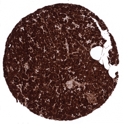
Mouse monoclonal / IgG1 1:100 – 1:200 Research Use Only Cytoplasmic and secreted. Human MSVA-601M Carboxypeptidase A1 (pancreatic); CPA1; Pancreatic Carboxypeptidase A1; Procarboxypeptidase A1 pancreatic Pancreas: A strong CPA1 staining of acinar cells should be seen. Adjacent structures can also be stained due to contamination artifacts. Colon: CPA1 immunostaining should not be seen. CPA1 is expressed on normal and neoplastic pancreatic acinar cells. Carboxypeptidase A1 (CPA1) is a zinc metalloprotease coded by the CPA1 gene located at 7q32.2. It is a 34,6kDa protein which is solely produced in the pancreas. It is involved in zymogen inhibition and was shown to preferentially cleave C-terminal branched-chain and aromatic amino acids from dietary proteins. Mutations of CPA1 gene have been linked to chronic pancreatitis. Elevated CPA1 serum protein levels have been described in patients with pancreatic cancer. [1] [1] Uhlig R et al. “Carboxypeptidase A1 (CPA1) Immunohistochemistry Is Highly Sensitive and Specific for Acinar Cell Carcinoma (ACC) of the Pancreas.” Am J Surg Pathol. 2022 Jan 1;46(1):97-104. PMID: 34889867. CPA1 staining pattern in Normal Tissues with antibody MSVA-601M (images are shown in our “Normal Tissue Gallery”) Brain Cerebrum Negative. Cerebellum Negative. Endocrine Tissues Thyroid Negative. Parathyroid Negative. Adrenal gland Negative. Pituitary gland Negative. Respiratory system Respiratory epithelium Negative. Lung Negative. Gastrointestinal Tract Salivary glands Negative. Esophagus Negative. Stomach Negative. Colon Negative. Duodenum Negative. Rectum Negative. Small intestine Negative. Liver Negative. Gallbladder Negative. Pancreas Strong cytoplasmic CPA1 staining in all acinar cells. CPA1 does not seem to be expressed in islet cells, intercalated ducts and excretory ducts – but all these cell types can show a variable intensity staining due to contamination artifacts caused by high levels of CPA1 in adjacent acinar cells. Genitourinary Kidney Negative. Urothelium Negative. Male genital Prostate Negative. Seminal vesicles Negative. Testis Negative. Epididymis Negative. Female ... CPA1 immunostaining is predominantly seen in acinus cell carcinomas of the pancreas. The TCGA findings on CPA1 RNA expression in different tumor categories have been summarized in the Human Protein Atlas. Acinar cell carcinoma of the pancreas showing strong CPA1 immunostaining in all tumor cells. Complete absence of CPA1 immunostaining in a ductal adenocarcinoma of the pancreas. Complete absence of CPA1 immunostaining in a neuroendocrine carcinoma of the pancreas. Cancer tissue gallery CPA1 (MSVA-601M) publication summary: Relevant publication: Uhlig et al. “Carboxypeptidase A1 (CPA1) Immunohistochemistry is Highly Sensitive and Specific for Acinar Cell Carcinoma (ACC) of the Pancreas” . Published in the American Journal of Surgical Pathology. 2022 Jan 1;46(1):97-104. A total of 12,274 tumors were analyzed from 131 different tumor categories by using the following protocol: Heat-induced antigen retrieval for 5 minutes in an autoclave at 121°C in pH7,8 Target Retrieval Solution buffer. MSVA-601M at a dilution of 1:150 at 37°C for 60 minutes. Visualization of bound antibody by the EnVision Kit (Dako, Agilent). This protocol was also used for all stainings depicted in our tumor and normal tissue galleries. A positive CPA1 immunostaining was seen in all 12 analyzed pancreatic acinar cell carcinomas but in none of 12263 tumors from 130 other tumor categories. The distribution of positive staining results is shown in an “organ-systematic” figure below (images based on dat... IHC users have different preferences on how the stains should look like. Some prefer high staining intensity of the target stain and even accept some background. Others favor absolute specificity and lighter target stains. Factors that invariably lead to more intense staining include higher concentration of the antibody and visualization tools, longer incubation time, higher temperature during incubation, higher temperature and longer duration of the heat induced epitope retrieval (slide pretreatment). The impact of the pH during slide pretreatment has variable effects and depends on the antibody and the target protein. All images and data shown here are obtained by the manual protocol described below. Protocols resulting in equivalent staining by using an automated immunostainer are described as well. Visualization of bound antibody by the EnVision Kit (Dako, Agilent) according to the manufacturer’s directions. Manual protocol Freshly cut sections should be used (less than 10 days bet... CPA1 expression has been shown to represent a specific feature of pancreatic acinar cell carcinoma. Further studies are needed to validate this findings. The prognostic relevance of different CPA1 expression levels should be investigated in pancreatic acinar cell carcinoma. Can elevated CPA1 serum levels already suggest acinar cell carcinoma There are two ways, how the specificity of antibodies can be documented for immunohistochemistry on formalin fixed tissues. These are: 1. comparison with a second independent method for target expression measurement across a large number of different tissue types (orthogonal strategy), and 2. Comparison with one or several independent antibodies for the same target and showing that all positive staining results are also seen with other antibodies for the same target (independent antibody strategy). For the antibody MSVA-601M specificity is documented by the strong concordance of the immunostaining with RNA expression data derived from the Human Protein Atlas (HPA) RNA-seq tissue dataset , the FANTOM5 project, and the Genotype-Tissue Expression (GTEx) project which are all summarized in the Human Protein Atlas (Tissue expression CPA1) . Immunostaining by using MSVA-601M was exclusively detected in the pancreas, the only organ with documented RNA expression. Moreover, no staining was see...