CBP (HMV319)
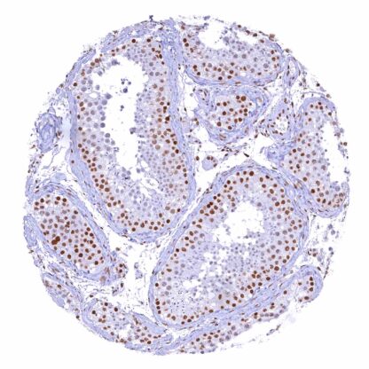
Recombinant Rabbit monoclonal / IgG 1:100 – 1:200 Research Use Only Cytoplasm,Nucleus Human HMV319 CREB binding protein,CBP,KAT3A,MKHK1,RSTS,RSTS1 Testis: A strong nuclear CBP staining should be seen in Sertoli and Leydig cells as well as in spermatogonia. Testis: The nuclear CBP staining intensity decreases during the maturation of spermatocytes while spermatids are completely CBP negative. CBP is a Histone acetyltransferase that acts as a master regulator of gene expression. CBP (CREB-binding protein) is a histone acetyltransferase coded by the CREBBP gene on chromosome 16p13.3. CBP co-operates closely with p300, another histone acetyltransferase with functional overlap and considerable sequence homology. Both p300 and CBP interact with numerous transcription factors to increase the expression of their target genes. They are thought to relax the chromatin structure at gene promoters through their intrinsic histone acetyltransferase activity, to recruit the basal transcriptional machinery including RNA polymerase II to the promoter, and to act as adaptor molecules. CBP and p300 are critical for normal embryonic development, as mice completely lacking either CBP or p300 protein, die at an early embryonic stage. That mice with a lack of one functional copy of both the CBP and p300 genes and thus having half of the normal amount of both CBP and p300 also die early in embryogenesis suggests that the total amount of CBP and p300 protein is critical for embryo d... Images describing the CBP staining pattern in normal tissues obtained by the antibody HMV319 are shown in our “ Normal Tissue Gallery ”. Brain Cerebrum Weak or absent CBP staining of neurons (perhaps due to overfixation, in tissues with prolonged fixation). Cerebellum Weak or absent CBP staining of a fraction of cells (perhaps due to overfixation, in tissues with prolonged fixation). Endocrine Tissues Thyroid Strong nuclear CBP positivity of all cells. Parathyroid Strong nuclear CBP positivity of all cells. Adrenal gland Strong nuclear CBP positivity of all cells. Pituitary gland CBP staining is weak or absent in a fraction of epithelial cells of the adenohypophysis. Respiratory system Respiratory epithelium Strong nuclear CBP positivity of all cells. Lung Strong nuclear CBP positivity of all cells. Gastrointestinal Tract Salivary glands Strong nuclear CBP positivity of all cells. Esophagus Strong nuclear CBP positivity of cells. In squamous epithelium, the CBP staining intensity decre... A nuclear CBP staining of various intensity is almost always seen in all kinds of cancers. Rarely, a CBP expression loss can occur in cancers, especially in malignant lymphomas. The TCGA findings on CBP RNA expression in different tumor categories have been summarized in the Human Protein Atlas. Adenocarcinoma with a complete lack of CBP staining in tumor cells Malignant peripheral nerve sheath tumor (MPNST) with strong CBP positivity of tumor cells Muscle-invasive urothelial carcinoma not showing any CBP immunostaining in tumor cells Cancer tissue gallery No data available at the moment IHC users have different preferences on how the stains should look like. Some prefer high staining intensity of the target stain and even accept some background. Others favor absolute specificity and lighter target stains. Factors that invariably lead to more intense staining include higher concentration of the antibody and visualization tools, longer incubation time, higher temperature during incubation, higher temperature and longer duration of the heat induced epitope retrieval (slide pretreatment). The impact of the pH during slide pretreatment has variable effects and depends on the antibody and the target protein. All images and data shown here and in our image galleries are obtained by the manual protocol described below. Other protocols resulting in equivalent staining are described as well. Manual protocol Freshly cut sections should be used (less than 10 days between cutting and staining). Heat-induced antigen retrieval for 5 minutes in an autoclave at 121°C in pH 7,8 Target ... The role of CBP as a therapeutic target needs to be further investigated. The predictive and prognostic role of CBP immunohistochemistry should be investigated. The role CBP in metabolic and neurologic disorders needs to be further evaluated. There are two ways how the specificity of antibodies can be documented for immunohistochemistry on formalin fixed tissues. These are: 1. Comparison with a second independent method for target expression measurement across a large number of different tissue types (orthogonal strategy), and 2. Comparison with one or several independent antibodies for the same target and showing that all positive staining results are also seen with other antibodies for the same target (independent antibody strategy). Orthogonal validation: For the antibody HMV319 specificity is supported by the good concordance of the immunostaining data with data from three independent RNA screening studies, including the Human Protein Atlas (HPA) RNA-seq tissue dataset, the FANTOM5 project, and the Genotype-Tissue Expression (GTEx) project, which are all summarized in the Human Protein Atlas (Tissue expression CBP) . In agreement with HMV319 immunostaining data, CBP RNA expression predominated in the bone marrow and lym...