Cytokeratin 5 (MSVA-605M)
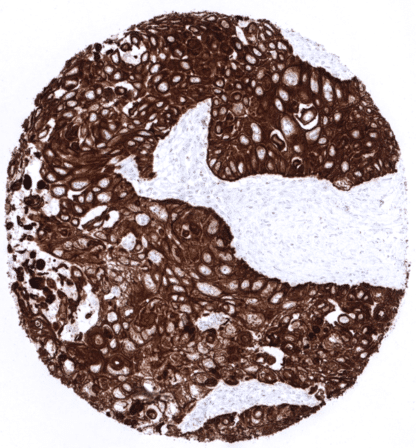
Recombinant Mouse monoclonal / IgG1 1:100 – 1:200 Research Use Only Cytoplasmic Human MSVA-605M 58kDa Cytokeratin; CK5; Cytokeratin-5; DDD1; Epidermolysis Bullosa Simplex 2 (EBS2); Keratin 5; Keratin, Type II Cytoskeletal 5; Keratin-5; KRT5; Type-II Cytoskeletal 5; Type-II keratin Kb5 Tonsil: A moderate to strong cytoplasmic KRT5 staining should be seen in virtually all squamous epithelial cells. Prostate: A strong cytoplasmic staining should be present in the majority of basal cells. Tonsil: Lymphatic cells must not show any KRT5 positivity. Cytokeratin 5 is expressed in basal cells and cancers derived from cytokeratin 5 expressing normal tissues. Cytokeratin 5 (CK5), also termed keratin 5 (KRT5) is a type II keratin protein encoded by the KRT5 gene on 12q13.13. It dimerizes with the type I keratin 14 and forms the intermediate filaments that primarily shape the cytoskeleton of basal epithelial cells. As other cytokeratins, KRT5 is part of the cytoskeletal scaffold within epithelial cells, which contributes to the cell architecture and provides the cells with the ability to withstand mechanical stress. Together with its partner protein keratin 14, KRT5 makes up for up to 25% of total cell protein in epidermal basal cells. Mutations of the KRT5 gene are leading to epidermolysis, an inherited skin blistering disease. The severity of the disease is linked to the position of the mutation within the protein. Expression of KRT5 (and 14) in cancer is indicating basal-like subtype which is linked to poor prognosis in several cancer types including breast and lung cancer. [1] [1] Völkel et al. “Cytokeratin 5 and cytokeratin 6 expressions... Cytokeratin 5 staining pattern in Normal Tissues with antibody MSVA-605M (images are shown in our “Normal Tissue Gallery”) Brain Cerebrum Negative. Cerebellum Negative. Endocrine Tissues Thyroid Negative Parathyroid Negative Adrenal gland Negative. Pituitary gland Negative. Respiratory system Respiratory epithelium Strong CK5 staining in basal cells of respiratory epithelium. Moderate CK5 staining inmyoepithelial cells and basal cells of excretory ducts of bronchial glands. Lung Negative. Gastrointestinal Tract Salivary glands Strong CK5 staining in myoepithelial cells and basal cells of excretory ducts. Esophagus Strong CK5 positivity of all squamous epithelial cell layers. Tendency to a slightly decreased staining towards the surface cell layers. Stomach Negative. Duodenum Negative. Small intestine Negative. Appendix Negative. Colon Negative. Rectum Negative. Liver Negative. Gallbladder Negative. Pancreas Negative. Genitourinary Kidney Negative. Urothelium In the urothelium, only the... KRT5 expression is seen in almost all squamous cell carcinomas irrespective of their origin and differentiation. KRT5 is also seen in almost all thymic epithelial tumors, >80% of epitheloid type malignant mesothelioma, >60% of urothelial carcinomas of the urinary bladder, about 40% of endometroid carcinomas of the endometrium or ovary, 25-35% of pancreatic adenocarcinomas, 15-20% of cholangiocarcinomas of the liver, and in other cancers. Detailed data on CK5 staining by MSVA-605M obtained from an analysis of 12,525 tumors from 120 different tumor types and subtypes have recently been published by Völkel et al. “Cytokeratin 5 and cytokeratin 6 expressions are unconnected in normal and cancerous tissues and have separate diagnostic implications” The TCGA findings on Cytokeratin 5 RNA expression in different tumor categories have been summarized in the Human Protein Atlas. Strong KRT5 immunostaining in a squamous cell carcinoma of the lung. Strong KRT5 expression in all tumor cells of a t... Cytokeratin 5 (MSVA-605M) publication summary: Relevant publication: Völkel et al. Cytokeratin 5 and cytokeratin 6 expressions are unconnected in normal and cancerous tissues and have separate diagnostic implications” Published in Virchows Arch. 2022 Feb;480(2) PMID: 34559291 In this study, 12’525 tumors were analyzed from 120 different tumor categories by using the following protocol: Heat-induced antigen retrieval for 5 minutes in an autoclave at 121°C in pH 9 Target Retrieval Solution buffer. MSVA-605M at a dilution of 1:150 at 37°C for 60 minutes. Visualization of bound antibody by the EnVision Kit (Dako, Agilent). This protocol was also used for all stainings depicted in our tumor and normal tissue galleries. At least one case with a positive CK5 immunostaining was seen in 62 (51.7%) and at least one case with a strong CK5 immunostaining was seen in 48 (40%) of 120 tumor categories. The distribution of positive staining results is shown in an “organ-systematic” (Figure 1) and in a... IHC users have different preferences on how the stains should look like. Some prefer high staining intensity of the target stain and even accept some background. Others favor absolute specificity and lighter target stains. Factors that invariably lead to more intense staining include higher concentration of the antibody and visualization tools, longer incubation time, higher temperature during incubation, higher temperature and longer duration of the heat induced epitope retrieval (slide pretreatment). The impact of the pH during slide pretreatment has variable effects and depends on the antibody and the target protein. Accordingly, multiple different protocols can generate identical staining results. All images and data shown here and in our image galleries are obtained by the manual protocol described below. Other protocols resulting in equivalent staining are described as well. Manual protocol Freshly cut sections should be used (less than 10 days between cutting and staining). Heat... Visualization of basal cells and myoepithelial cells in multicolor IHC approaches. The potential diagnostic utility of KRT5 expression analysis (as compared to KRT5/6 analysis) needs to be investigated. The clinical significance of KRT5 expression needs to be evaluated in tumor types containing significant subgroups of KRT5 positive and negative tumors. Specificity of MSVA-605M is documented by strong positive staining in cell types that are well documented to express KRT5 such as squamous epithelia of all origins and basal cells in the prostate in combination with the absence of staining in all tissues known to not express KRT5 including tissues notorious for non-specific IHC background such as kidney, colonic mucosa, and epidermis. In a recent publication, the specificity of MSVA-605M was demonstrated in a comparison vs another independent antibody. Völkel et al. “Cytokeratin 5 and cytokeratin 6 expressions are unconnected in normal and cancerous tissues and have separate diagnostic implications” Normal tissue gallery