Cytokeratin 6 (MSVA-606R)
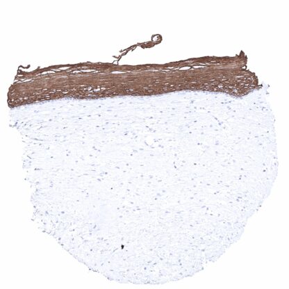
Recombinant Rabbit monoclonal / IgG1 1:100 – 1:200 Research Use Only Cytoplasmic Human MSVA-606R CK6A, CK6B, CK6C, CK6D, CK6E, Keratin Type II Cytoskeletal 6A, Keratin Type II Cytoskeletal 6B, Keratin Type II Cytoskeletal 6C, Keratin Type II Cytoskeletal 6D, Keratin Type II Cytoskeletal 6E, KRT6, KRT6A, KRT6B, KRT6C, KRT6D, KRT6E Tonsil: A strong CK6 staining should be seen in all epithelial cells of the surface epithelium except the basal cell layer where staining should be absent or weak. Tonsil: CK6 staining should be absent in lymphocytes and all other mesenchymal cells. Cytokeratin 6 (CK6) or keratin 6 (KRT6) is a basic high molecular weight Type II cytokeratin. It is an integral part of the cytoskeleton of various mostly squamous epithelial cell types in various organs. There are 3 keratin 6 isoforms which are all coded by neighboring genes located within the type II keratin gene cluster on chromosome 12q. The Keratins 6A, 6B and 6C genes consist of 9 exons and share more than 99% similarity in their DNA coding sequences. Most keratins form heteropolymers consisting of a type I and a type II keratin. All keratin 6 isoforms pair with keratin 16 and/or keratin 17. Keratin 6A has antimicrobial properties. Mutations of keratin 6 lead to the PC-K6A subtype of pachyonychia congenita with structural abnormalities of the nails, the epidermis of the palms and soles, and oral epithelia. [1] [1] Völkel et al. “Cytokeratin 5 and cytokeratin 6 expressions are unconnected in normal and cancerous tissues and have separate diagnostic implications” Virchows Archiv p... Cytokeratin 6 staining pattern in Normal Tissues with antibody MSVA-606R (images are shown in our “Normal Tissue Gallery”) Brain Cerebrum Negative. Cerebellum Negative. Endocrine Tissues Thyroid Negative. Parathyroid Negative. Adrenal gland Negative. Pituitary gland Negative. Respiratory system Respiratory epithelium Moderate to strong CK6 staining of basal cells and sometimes also ciliated cells of respiratory epithelium. Moderate to strong positivity also in intercalated ducts of bronchial glands. Lung Negative. Gastrointestinal Tract Salivary glands Moderate to strong CK6 positivity in intercalated ducts. Esophagus Strong CK6 positivity in the squamous epithelium but the basal cell layer is negative. Stomach Negative. Duodenum Negative. Small intestine Negative. Appendix Negative. Colon Negative. Rectum Negative. Liver Negative. Gallbladder Negative. Pancreas Negative. Genitourinary Kidney Negative. Urothelium Negative. Male genital Prostate Negative. Seminal vesicles Negative. Test... CK6 expression is seen in almost all squamous cell carcinomas irrespective of their origin. CK6 is also seen in 30-50% of urothelial carcinomas of the urinary bladder, 30-50% of pancreatic adenocarcinomas, 20-30% of endometroid carcinomas of endometrium and ovary, 10-20% of cholangiocarcinomas of the liver, 10-20% of adenocarcinomas of the lung, and in other cancers. KRT6 immunostaining can usually be seen in only a few cells of basaliomas. Detailed data on CK6 staining by MSVA-606R obtained from an analysis of 12,898 tumors from 120 different tumor types and subtypes have recently been published by Völkel et al. “Cytokeratin 5 and cytokeratin 6 expressions are unconnected in normal and cancerous tissues and have separate diagnostic implications” The TCGA findings on Cytokeratin 6 RNA expression in different tumor categories have been summarized in the Human Protein Atlas. Squamous cell carcinoma of the oral cavity with strong CK6 immunostaining. CK6 negative breast cancer Diffuse, str... CK6 staining in tumors with antibody MSVA-606R. Data from the publication “ Cytokeratin 5 and cytokeratin 6 expressions are unconnected in normal and cancerous tissues and have separate diagnostic implications ” published by Völkel et al. in Virchows Archive September 2021. Summarized in own graphics: CK6 staining in tumors with antibody MSVA-606R 2. CK6 staining in tumors with antibody MSVA-606R ranked by positivity. Figure 3. Clinico-pathological associations described by Völkel et al. (p-value) IHC users have different preferences on how the stains should look like. Some prefer high staining intensity of the target stain and even accept some background. Others favor absolute specificity and lighter target stains. Factors that invariably lead to more intense staining include higher concentration of the antibody and visualization tools, longer incubation time, higher temperature during incubation, higher temperature and longer duration of the heat induced epitope retrieval (slide pretreatment). The impact of the pH during slide pretreatment has variable effects and depends on the antibody and the target protein. Accordingly, multiple different protocols can generate identical staining results. All images and data shown here and in our image gallery are obtained by the manual protocol described below. Other protocols resulting in equivalent staining are described as well. Manual protocol Freshly cut sections should be used (less than 10 days between cutting and staining). Heat-i... The potential diagnostic utility of KRT6 expression analysis (as compared to KRT5/6 analysis) needs to be investigated. The clinical significance of KRT6 expression needs to be evaluated in tumor types containing significant subgroups of KRT6 positive and negative tumors. Specificity of MSVA-606R is documented by strong positive staining in cell types that are well documented to express CK6 such as squamous epithelia of all origins and absence of staining in all tissues known to not express CK6 including tissues notorious for non-specific IHC background such as kidney, colonic mucosa, and epidermis. In a recent publication, the specificity of MSVA-606R was demonstrated in a comparison vs another independent antibody. Völkel et al. “Cytokeratin 5 and cytokeratin 6 expressions are unconnected in normal and cancerous tissues and have separate diagnostic implications” Normal tissue gallery