Cytokeratin 7 (MSVA-607R)
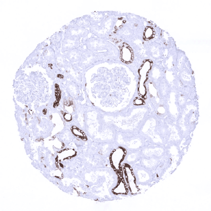
Recombinant Rabbit monoclonal / IgG 1:100 – 1:200 Research Use Only Cytoplasmic Human MSVA-607R CK-7, K2C7, Keratin 55K Type II Cytoskeletal, Keratin Simple Epithelial Type 1 K7, Keratin Type II Cytoskeletal 7, Krt2-7, KRT7, Sarcolectin, SCL, Type II Mesothelial Keratin K7, Type-II Keratin Kb7 Pancreas: CK7 staining is at least weak to moderate cytoplasmic intercalating ducts and strong in large pancreatic ducts. Pancreas: CK7 staining should be absent in all acinar cells. Cytokeratin 7 (CK7), also termed keratin 7 (KRT7) is a basic high molecular weight Type II cytokeratin encoded by the KRT7 gene located12q12-q13. It forms intermediate filaments that primarily shape the cytoskeleton of specific epithelial, mostly glandular cells. In these cells, CK7 is part of the cytoskeletal scaffold which contributes to the cell architecture and provides the cells with the ability to withstand mechanical stress. Because of its differential expression in carcinomas from different origins, CK7 immunohistochemistry can be used as an aide for defining the origin of cancer tissues. Cytokeratin 7 (CK7), also termed keratin 7 (KRT7) is a basic high molecular weight Type II cytokeratin encoded by the KRT7 gene located 12q12-q13 . It forms intermediate filaments that primarily shape the cytoskeleton of specific epithelial, mostly glandular cells. In these cells, CK7 is part of the cytoskeletal scaffold which contributes to the cell architecture and provides the cells with the ability to withstand mechanical stress. Because of its differential expression in carcinomas from different origins, CK7 immunohistochemistry can be used as an aide for defining the origin of cancer tissues. Cytokeratin 7 staining pattern in Normal Tissues with antibody MSVA-607R (images are shown in our “Normal Tissue Gallery”) Brain Cerebrum Negative. Cerebellum Negative. Endocrine Tissues Thyroid Strong CK7 staining of follicular epithelial cells. Parathyroid Weak to moderate CK7 staining of a fraction of epithelial cells. Adrenal gland Negative. Pituitary gland Moderate CK7 staining of a small fraction of epithelial cells of the adenohypophysis (not in all samples). Respiratory system Respiratory epithelium Strong CK7 staining of suprabasal cells. Weak staining of basal cells. Lung Moderate CK7 staining of pneumocytes. Strong CK7 staining of mucinous cells, serous cells, and excretory ducts of bronchial glands. Gastrointestinal Tract Salivary glands Strong CK7 staining of mucinous cells, serous cells, and excretory ducts. Esophagus Usually negative. Faint staining of few cells can occur. Stomach Moderate CK7 staining of most surface epithelial cells. Duodenum CK7 staining in few scatte... KRT7 is expressed in a wide range of tumors, most of which are derived from KRT7 positive precursor cells. The highest frequency of KRT7 positivity is seen in carcinomas of the ovary, thyroid, urothelium, breast, and endometrium, chromophobe and papillary kidney cancer, adenocarcinomas of the pancreas, lung, esophagus, and the stomach as well as in mesothelioma. Many other tumor types can – at least occasionally – show KRT7 expression. The TCGA findings on Cytokeratin 7 RNA expression in different tumor categories have been summarized in the Human Protein Atlas. Gastric cancer (intestinal type) with strong KRT7 immunostaining of tumor cells. Invasive breast cancer of no special type with strong KRT7 immunostaining in all tumor cells. Clear cell renal cell carcinoma with weak focal KRT7 positivity. Cancer tissue gallery Cytokeratin 7 (MSVA-607R) publication summary Relevant publication: Dum et al.: “Cytokeratin 7 and cytokeratin 20 expression in cancer: A tissue microarray study on 15,424 cancers” Exp Mol Pathol 2022 Apr 4; 126:104762 Online ahead of print A total of 14’110 tumors from 120 different tumor categories were successfully analyzed for CK 7 by using the following protocol: Heat-induced antigen retrieval for 5 minutes in an autoclave at 121°C in pH 9 Target Retrieval Solution buffer. MSVA- 620R at a dilution of 1:150 at 37°C for 60 minutes. Visualization of bound antibody by the EnVision Kit (Dako, Agilent). This protocol was also used for all CK7 immunostainings depicted in our tumor and normal tissue galleries. At least one case with a positive CK7 immunostaining was seen in 89 (74.2%) and at least one case with a strong CK7 staining was seen in 72 (60%) of 120 tumor categories. The distribution of positive staining results is shown in an “organ-systematic” (Figure 1) and in a “ranking or... IHC users have different preferences on how the stains should look like. Some prefer high staining intensity of the target stain and even accept some background. Others favor absolute specificity and lighter target stains. Factors that invariably lead to more intense staining include higher concentration of the antibody and visualization tools, longer incubation time, higher temperature during incubation, higher temperature and longer duration of the heat induced epitope retrieval (slide pretreatment). The impact of the pH during slide pretreatment has variable effects and depends on the antibody and the target protein. Accordingly, multiple different protocols can generate identical staining results. All images and data shown here and in our image galleries are obtained by the manual protocol described below. Other protocols resulting in equivalent staining are described as well. Manual protocol Freshly cut sections should be used (less than 10 days between cutting and staining). Heat... As the literature is partly confusing, the diagnostic utility of KRT7 expression analysis (in combination with KRT20 analysis) should be investigated in a large cohort of tumors from different entities. The biologic/clinical significance of aberrant CK7 expression in cancers needs to be evaluated (for example: what are the specific properties of CK7 negative pancreatic cancers or CK7 positive colorectal cancers?) Specificity of MSVA-607R is documented by strong positive staining in cell types that are well documented to express CK7 such as epithelial cells of various glandular tissues and absence of staining in all tissues known to not express CK7 including tissues notorious for non-specific IHC background such as proximal tubules of the kidney, colon mucosa, and the skin. Normal tissue gallery