Cytokeratin 13 (MSVA-613M)
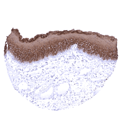
Mouse monoclonal / IgG1 1:100 – 1:200 Research Use Only Cytoplasmic Human MSVA-613M CK13; Cytokeratin-13; Keratin Type I Cytoskeletal 13; Keratin-13; KRT13; Type I Cytoskeletal 13; WSN2 Tonsil: All squamous epithelial cells of the surface squamous epithelium (except the basal layer) and a fraction of squamous cells of the crypts should show strong KRT13 staining. Tonsil: All lymphocytes and blood vessels must not show any KRT13 staining. Cytokeratin 13 is expressed in non-keratinizing squamous epithelial cells. Cytokeratin 13 (CK13), also termed keratin 13 (KRT13) is a type I acidic high molecular weight keratin protein encoded by the KRT13 gene located at 17q21. It dimerizes with the type I keratin 4 and forms intermediate filaments that primarily shape the cytoskeleton of specific epithelial cells. In these cells, KRT13 is part of the cytoskeletal scaffold which contributes to the cell architecture and provides the cells with the ability to withstand mechanical stress. Mutations in CK10 have been linked to the autosomal dominant disorder “White Sponge Nevus”. Cytokeratin 13 staining pattern in Normal Tissues with antibody MSVA-613M (images are shown in our “Normal Tissue Gallery”) Brain Cerebrum Negative. Cerebellum Negative. Endocrine Tissues Thyroid Negative. Parathyroid Negative. Adrenal gland Negative. Pituitary gland Negative. Respiratory system Respiratory epithelium Very few KRT13 positive basal cells can occur. Lung Negative. Gastrointestinal Tract Salivary glands Very few KRT13 positive individual cells or small groups of cells can occur in the epithelium. Focal staining of cells and groups of cells in excretory ducts of salivary glands Esophagus Strong CK13 staining of all supra-basal cell layers of non-keratinizing squamous epithelium. Stomach Negative. Duodenum Negative. Small intestine Negative. Appendix Negative. Colon Negative. Rectum Negative. Liver Negative. Gallbladder Faint staining of few gall bladder epithelial cells in a case with inflammation. Pancreas Negative. Genitourinary Kidney Negative. Urothelium Strong CK13 st... KRT13 immunostaining almost exclusively occurs in squamous cell carcinomas of various sites of origin. At frequencies <10% KRT13 expression can also be seen in adenocarcinomas of the pancreas, stomach, ovary, lung, endometrium, breast, esophagus, and the prostate, as well as in nonseminomatous germ cell tumors. The TCGA findings on Cytokeratin 13 RNA expression in different tumor categories have been summarized in the Human Protein Atlas. Esophageal squamous cell carcinoma with strong diffuse KRT13 positivity. Non-invasive papillary urothelial carcinoma (pTa) showing strong diffuse KRT13 positivity. KRT13 negative colorectal adenocarcinoma. Cancer tissue gallery Cytokeratin 13 (MSVA-613M) publication summary Relevant publication: Lennartz et al. “Cytokeratin 13 (CK13) expression in cancer: a tissue microarray study on 10,439 tumors” Published in the Journal of Pathology, Microbiology and Immunology – the APMIS journal 2022 Oct 21. doi: 10.1111/apm.13280. Epub ahead of print. PMID: 36269681. A total of 9,156 tumors from 131 different tumor categories were successfully analyzed by using the following protocol: Heat-induced antigen retrieval for 5 minutes in an autoclave at 121°C in pH 7,8 Target Retrieval Solution buffer. MSVA-613M at a dilution of 1:150 at 37°C for 60 minutes. Visualization of bound antibody by the EnVision Kit (Dako, Agilent). This protocol was also used for all stainings depicted in our tumor and normal tissue galleries. Overall, 42 (32,1%) of 137 tumor categories showed detectable CK13 staining in at least one case and 24 (18,3%) tumor categories included at least one case with strong CK13 positivity. The highest rate of pos... IHC users have different preferences on how the stains should look like. Some prefer high staining intensity of the target stain and even accept some background. Others favor absolute specificity and lighter target stains. Factors that invariably lead to more intense staining include higher concentration of the antibody and visualization tools, longer incubation time, higher temperature during incubation, higher temperature and longer duration of the heat induced epitope retrieval (slide pretreatment). The impact of the pH during slide pretreatment has variable effects and depends on the antibody and the target protein. Accordingly, multiple different protocols can generate identical staining results. All images and data shown here and in our image galleries were obtained by the manual protocol described below. Other protocols resulting in equivalent staining are described as well. Manual protocol Freshly cut sections should be used (less than 10 days between cutting and staining). Hea... The diagnostic utility of KRT713 expression analysis should be investigated in a large cohort of tumors from different entities. The diagnostic utility of increased KRT13 expression in the skin is not known. The diagnostic utility of decreased KRT13 expression in non-keratinizing squamous epithelium of ectocervix, esophagus, oral cavity and lip is not known. The prognostic role of KRT13 expression in squamous cell carcinoma is unknown. Utility of MSVA-613M is documented by a staining pattern that exactly matches the data summarized in the protein atlas. A strong positive KRT13 staining is almost exclusively seen in non-keratinizing squamous epithelial cells but not in the skin. In addition, all tissues known to not express KRT13 including those notorious for non-specific IHC background such as kidney and colonic mucosa are completely KRT13 negative. Normal tissue gallery