Cytokeratin 14 (MSVA-614R)
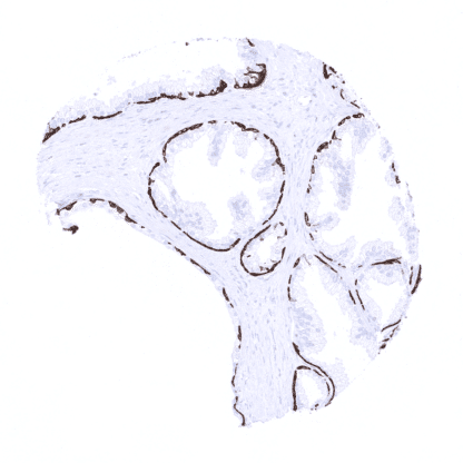
Recombinant Rabbit monoclonal / IgG 1:50 – 1:100 Research Use Only Cytoplasmic Human MSVA-614R CK-14; Dowling Meara; ebs3; ebs4; Epidermolysis Bullosa Simplex; Keratin Type I Cytoskeletal 14; Koebner; NFJ Tonsil: A strong KRT14 immunostaining should be seen in the surface and crypt epithelium. Tonsil: KRT14 immunostaining should be absent in all non-epithelial cells. Cytokeratin 14 is expressed in basal and myoepithelial cells of the prostate and the breast. Cytokeratin 14 (CK14), also termed keratin 14 (KRT14) is a 50 kDa type I acidic keratin protein encoded by the KRT14 gene at 17q21.2. KRT14 is part of the cytoskeletal scaffold within epithelial cells, which contributes to the cell architecture and provides the cells with the ability to withstand mechanical stress. Keratin 14 is usually found as a heterodimer with type II keratin 5. Mutations in KRT14 are associated with epidermolysis bullosa simplex and dermatopathia pigmentosa reticularis. In the skin and in non-keratinizing squamous epithelium all cell layers usually show a strong KRT14 staining, but staining is sometimes weaker or even absent in the more superficial cell layers. Strong KRT14 positivity is also seen in sebaceous glands and hair follicles, epithelial cells including corpuscles of Hassall’s in the thymus, tonsil crypt epithelium, basal cells of the prostate and respiratory epithelium as well as in Amnion and chorion cells of the placenta. In salivary glands, KRT14 is strongly expressed in myoepithelial cells, basal cells and a fraction of ductal cells of excretory ducts. KRT14 immunostaining is absent in lung, liver, pancreas, epididymis, testis, urothelium, gastrointestinal epithelial cells, Brunner glands, fallopian tube, trophoblastic cells of the placenta, adrenal gland, parathyroid gland, brain, adeno- and neurohypophysis, spleen, lymph node, all hematopoetic cell types, and all mesenchymal tissues. These findings are largely consistent with the RNA... KRT14 expression is seen in the majority of squamous cell carcinomas irrespective of their origin and differentiation. KRT14 is also seen in almost all thymic epithelial tumors, in a large fraction of epitheloid type malignant mesothelioma and urothelial carcinomas, and – at lower frequency – also in other cancers. The TCGA findings on Cytokeratin 14 RNA expression in different tumor categories have been summarized in the Human Protein Atlas. Squamous cell carcinoma of the esophagus with strong, predominantly basal KRT14 immunostaining. KRT14 negative clear cell renal cell carcinoma. Squamous cell carcinoma of the oral cavity with strong KRT14 positivity in all tumor cells. Cancer tissue gallery No data available at the moment IHC users have different preferences on how the stains should look like. Some prefer high staining intensity of the target stain and even accept some background. Others favor absolute specificity and lighter target stains. Factors that invariably lead to more intense staining include higher concentration of the antibody and visualization tools, longer incubation time, higher temperature during incubation, higher temperature and longer duration of the heat induced epitope retrieval (slide pretreatment). The impact of the pH during slide pretreatment has variable effects and depends on the antibody and the target protein. All images and data shown here are obtained by the manual protocol described below. Other protocols resulting in equivalent staining are described as well. Manual protocol Freshly cut sections should be used (less than 10 days between cutting and staining). Heat-induced antigen retrieval for 5 minutes in an autoclave at 121°C in pH 7,8 Target Retrieval Solution buffer. ... The diagnostic utility of KRT14 expression analysis should be investigated in a large cohort of tumors from different entities. The prognostic role of KRT14 expression needs to be evaluated for various tumor types. Identification of basal and myoepithelial cell types in the prostate, breast and salivary glands (in multicolor immunofluorescence) There are two ways how the specificity of antibodies can be documented for immunohistochemistry on formalin fixed tissues. These are: 1. comparison with a second independent method for target expression measurement across a large number of different tissue types (orthogonal strategy), and 2. Comparison with one or several independent antibodies for the same target and showing that all positive staining results are also obtained with other antibodies for the same target (independent antibody strategy). For the antibody MSVA-614R specificity is documented by the strong concordance of the immunostaining with RNA expression data derived from the Human Protein Atlas (HPA) RNA-seq tissue dataset , the FANTOM5 project, and the Genotype-Tissue Expression (GTEx) project which are all summarized in the Human Protein Atlas (Tissue expression Cytokeratin 14) These RNA data only highlight organs covered by squamous epithelium and salivary glands as KRT14 expressing tissues. Immunostaining by using ...