Cytokeratin 19 (MSVA-119M)
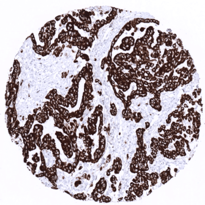
Mouse monoclonal / IgG1 1:100 – 1:200 Research Use Only Cytoplasmic Human MSVA-119M k19; k1cs; Keratin 19 Keratin Type i 40kD; krt19 Colon: A strong immunostaining is seen in all epithelial cells. Tonsil: A moderate to strong immunostaining occurs in the basal cell layer of the squamous epithelium of the tonsil surface. Colon: All non-epithelial cells are CK19 negative. Tonsil: All non-basal squamous epithelial cells and all non-epithelial cells are CK19 negative. Cytokeratin 19 is highly expressed in many tumor types but virtually absent in hepatocellular carcinoma. Cytokeratin 19 (CK19) is an acidic cytokeratin encoded by the KRT19 gene located in a keratin gene cluster at the human chromosome 17q12-q21. With a molecular mass of only 40kD, CK19 is the smallest member of the type 1 family of human keratins. While type 1 acidic cytokeratins typically form dimers with a type 1 basic cytokeratin, CK19 is an exception from this rule. However, like other epithelial cytokeratins, CK19 functions as a cytoskeletal scaffold protein to shape and strengthen the epithelial cell layer. Immunohistochemical analysis of cytokeratin 19 is often applied in diagnostic pathology because the protein is abundantly expressed in the majority of epithelial tumors. [1] [1] Menz et al. “Diagnostic and prognostic impact of cytokeratin 19 expression analysis in human tumors: a tissue microarray study of 13,172 tumors” Human Pathology: Volume 115, September 2021 Cytokeratin 19 staining pattern in Normal Tissues with antibody MSVA-119M (images are shown in our “Normal Tissue Gallery”) Brain Cerebrum Negative. Cerebellum Negative. Endocrine Tissues Thyroid Negative. Parathyroid Negative. Adrenal gland Negative. Pituitary gland Negative. Respiratory system Respiratory epithelium A moderate to strong Upk1b staining occurs in a fraction of cells. Lung Gastrointestinal Tract Salivary glands A variable (weak to strong) Upk1b positivity can sometimes occur in some serous cells in salivary glands. Esophagus Usually negative. Scattered Upk1b positive cells can occur. Stomach A moderate to strong Upk1b staining is seen in superficial epithelial cells and parietal cells. Colon Negative. Duodenum Negative. Rectum Negative. Small intestine Negative. Liver A variable (weak to strong) cytoplasmic and membranous Upk1b positivity can sometimes occur in intrahepatic bile ducts. Hepatocytes are negative. Gallbladder A variable (weak to strong) cytoplasmic and mem... Cytokeratin 19 is almost always expressed in various important cancers such as in adenocarcinomas of the stomach, esophagus, and the colorectum, breast cancers, ovarian cancers, endometrium carcinoma cholangiocarcinomas, and urothelial carcinomas. CK19 immunostaining is usually absent in hepatocellular carcinoma, adrenal adenomas, seminomas, hematological malignancies, and most mesenchymal tumors. Detailed data on Cytokeratin 19 staining by MSVA-119M obtained from an analysis of 13,172 tumors from 122 different tumors and subtypes have recently been published by Menz et al. “Diagnostic and prognostic impact of cytokeratin 19 expression analysis in human tumors: a tissue microarray study of 13,172 tumors” The TCGA findings on Cytokeratin 19 RNA expression in different tumor categories have been summarized in the Human Protein Atlas. Chromophobe renal cell carcinoma with intense cytokeratin 19 positivity. Papillary carcinoma of the thyroid with strong cytokeratin 19 immunostaining. Vul... Cytokeratin 19 (MSVA-119M) publication summary: Menz et al.: “Diagnostic and prognostic impact of cytokeratin 19 expression analysis in human tumors: a tissue microarray study of 13,172 tumors” Human Pathology: Volume 115, September 2021 A total of 13,172 tumors were analyzed from 122 different tumor categories by using the following protocol: Heat-induced antigen retrieval for 5 minutes in an autoclave at 121°C in pH7,8 Target Retrieval Solution buffer. MSVA-119M at a dilution of 1:150 at 37°C for 60 minutes. Visualization of bound antibody by the EnVision Kit (Dako, Agilent). This protocol was also used for all stainings depicted in our tumor and normal tissue galleries. At least one case with a positive cytokeratin 19 immunostaining was seen in 89 (73%) and at least one case with a strong cytokeratin 19 immunostaining was seen in 87 (71,3%) of 122 tumor categories. The distribution of positive staining results is shown “organ-systematic” and in a “ranking order” figure below (image... IHC users have different preferences on how the stains should look like. Some prefer high staining intensity of the target stain and even accept some background. Others favor absolute specificity and lighter target stains. Factors that invariably lead to more intense staining include higher concentration of the antibody and visualization tools, longer incubation time, higher temperature during incubation, higher temperature and longer duration of the heat induced epitope retrieval (slide pretreatment). The impact of the pH during slide pretreatment has variable effects and depends on the antibody and the target protein. All images and data shown here and in our product galleries are obtained by the manual protocol described below. Other protocols resulting in equivalent staining are described as well. Manual protocol Freshly cut sections should be used (less than 10 days between cutting and staining). Heat-induced antigen retrieval for 5 minutes in an autoclave at 121°C in pH 7,8 Targe... The diagnostic utility of KRT19 expression analysis should be investigated in a large cohort of tumors from different entities. Specificity of MSVA-119M is documented by strong positive staining in cell types that are well documented to express KRT19 such as a large variety of epithelial cell types and absence of staining in all tissues known to not express KRT19 including hepatocytes, adrenal gland, testis, mesenchymal tissues, lymphatic tissues and the brain. Normal tissue gallery