Cytokeratin 20 (MSVA-620R)
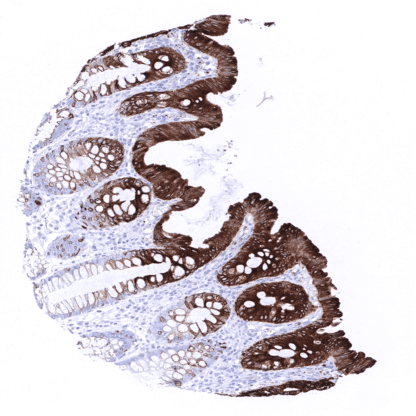
Recombinant Rabbit monoclonal / IgG 1:100 – 1:200 Research Use Only Cytoplasmic Human MSVA-620R CK20; Cytokeratin-20; K20; KA20; Keratin 20; keratin 20, type I; Keratin type I cytoskeletal 20; Keratin-20; KRT20 Colon mucosa: Staining should be strong at the surface and at least moderate at the basis of krypts. Tonsil: All cells must stain negative. Cytokeratin 20 (CK20) also termed keratin 20 (KRT20) is an acidic low molecular weight Type I cytokeratin encoded by the KRT20 gene located at 17q21.2. It forms intermediate filaments that primarily shape the cytoskeleton of specific epithelial cells, mainly of the gastrointestinal tract. CK20 expression is often retained in carcinomas derived from the gastrointestinal tract. CK20 immunohistochemistry can thus be used for defining the origin of cancer tissues. Because of a somewhat complementary staining pattern with CK7, CK20 IHC is often used together with CK7. Cytokeratin 20 (CK20) also termed keratin 20 (KRT20) is an acidic low molecular weight Type I cytokeratin encoded by the KRT20 gene located at 17q21.2. It forms intermediate filaments that primarily shape the cytoskeleton of specific epithelial cells, mainly of the gastrointestinal tract. CK20 expression is often retained in carcinomas derived from the gastrointestinal tract. CK20 immunohistochemistry can thus be used for defining the origin of cancer tissues. Because of a somewhat complementary staining pattern with CK7, CK20 IHC is often used together with CK7. Cytokeratin 20 staining pattern in Normal Tissues with antibody MSVA-620R (images are shown in our “Normal Tissue Gallery”) Brain Cerebrum Negative. Cerebellum Negative. Endocrine Tissues Thyroid Negative. Parathyroid Negative. Adrenal gland Negative. Pituitary gland Negative. Respiratory system Respiratory epithelium Negative. Lung Negative. Gastrointestinal Tract Salivary glands Negative. Esophagus Negative. Stomach Strong CK20 positivity in surface epithelial cells. CK20 staining is absent in the deep glands. Duodenum Strong CK20 staining of surface epithelial cells. The fraction of positive cells and the staining intensity decreases towards the base of glands/crypts. Small intestine Strong CK20 staining of surface epithelial cells. The fraction of positive cells and the staining intensity decreases towards the base of glands/crypts. Appendix Strong CK20 staining of surface epithelial cells. The fraction of positive cells and the staining intensity decreases towards the base of cryp... CK20 is commonly expressed in colorectal adenocarcinoma (95%), Merkel cell carcinoma, and mucinous ovarian cancer (60-70%) In Merkel cell carcinoma a characteristic dot like staining pattern is typically seen. In urothelial carcinoma, CK20 staining is more commonly seen in dysplasia, carcinoma in situ and non-invasive papillary cancer than in muscle invasive disease. CK20 staining occurs in 20-40% of adenocarcinomas of stomach, pancreas, and esophagus. At lower frequency CK20 expression can be seen in various other tumors especially adenocarcinomas. The TCGA findings on Cytokeratin 20 RNA expression in different tumor categories have been summarized in the Human Protein Atlas. Colorectal adenocarcinoma with strong CK20 immunostaining. Gastric adenocarcinoma with strong CK20 positivity of all tumor cells. Strong CK20 positivity of all tumor cells of a diffusely growing adenocarcinoma of the stomach. Cancer tissue gallery Cytokeratin 20 (MSVA-620R) publication summary Relevant publication: Dum et al.: “Cytokeratin 7 and cytokeratin 20 expression in cancer: A tissue microarray study on 15,424 cancers” Exp Mol Pathol 2022 Apr 4; 126:104762 Online ahead of print A total of 14’110 tumors from 120 different tumor categories were successfully analyzed for CK 20 by using the following protocol: Heat-induced antigen retrieval for 5 minutes in an autoclave at 121°C in pH 9 Target Retrieval Solution buffer. MSVA-620R at a dilution of 1:150 at 37°C for 60 minutes. Visualization of bound antibody by the EnVision Kit (Dako, Agilent). This protocol was also used for all CK20 immunostainings depicted in our tumor and normal tissue galleries. At least one case with a positive cytokeratin 20 staining was seen in 43 (35.8%) and at least one case with a strong cytokeratin 20 positivity was seen in 31 (25.8%) of 120 tumor categories. The distribution of positive staining results is shown in an “organ-systematic” (Figure 1)... IHC users have different preferences on how the stains should look like. Some prefer high staining intensity of the target stain and even accept some background. Others favor absolute specificity and lighter target stains. Factors that invariably lead to more intense staining include higher concentration of the antibody and visualization tools, longer incubation time, higher temperature during incubation, higher temperature and longer duration of the heat induced epitope retrieval (slide pretreatment). The impact of the pH during slide pretreatment has variable effects and depends on the antibody and the target protein. All images and data shown here and in our image galleries are obtained by the manual protocol described below. Other protocols resulting in equivalent staining are described as well. Manual protocol Freshly cut sections should be used (less than 10 days between cutting and staining). Heat-induced antigen retrieval for 5 minutes in an autoclave at 121°C in pH 9 Target Re... As the literature is partly confusing, the diagnostic utility of KRT20 expression analysis (in combination with KRT7 analysis) should be investigated in a large cohort of tumors from different entities. The biologic/clinical significance of aberrant KRT20 expression in cancers should be evaluated (for example: what are the specific properties of KRT20 negative colorectal cancers or of CK20 positive prostate cancers?) Specificity of MSVA-620R is documented by strong positive staining in cell types that are well documented to express CK20 such as surface epithelium of the stomach and colorectal mucosa and absence of staining in all tissues known to not express CK20 including tissues notorious for non-specific IHC background such as kidney and brain. Normal tissue gallery