DOG1 (MSVA-201M)
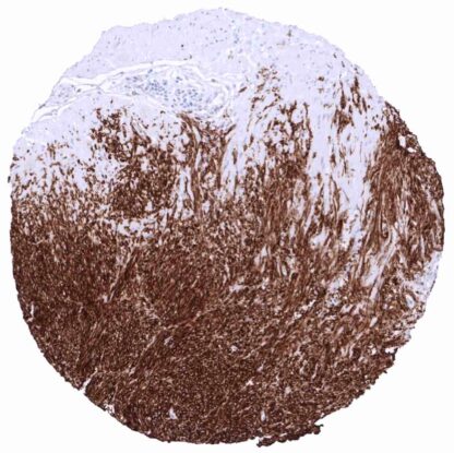
mouse monoclonal / IgG1 1:100 – 1:200 Research Use Only Cell Surface and Cytoplasm Human MSVA-201M Anoctamin 1; Calcium Activated Chloride Channel; Discovered On Gastrointestinal Stromal Tumors Protein 1; TAOS2; ORAOV2; TMEM16A Appendix: An at least moderate, predominantly membranous staining should be seen in Cajal cells in the muscular wall and a weak membranous staining should be seen in columnar epithelial cells in the basal part of the crypts of the appendix. Appendix: Staining should be absent in the majority of cells in the muscular wall and in epithelial cells located at the surface. DOG1 (Discovered on GIST1) was named after its discovery in gastrointestinal stroma tumors (GIST). The 111 KDa DOG1 protein (syn. TMEM16A, Anoctamin-1) is encoded by the ANO1 gene located at 11q13 and acts as a voltage-gated calcium-activated chloride and bicarbonate channel. In human tissues, DOG1 is normally expressed by the gastrointestinal interstitial cells of Cajal where it triggers epithelial chloride secretion that is required for intestinal motility. As the cells of Cajal give rise to gastrointestinal stroma tumors, retained expression of DOG1 is an important diagnostic feature of this tumor type. DOG1 exerts a direct impact ontumor growth in cancers through its regulation of EGFR signaling and pathway modulators like MAPK and protein kinase B. ANO1 blockers like niflumic acid have been shown to block slow waves, which produce motility, in the human intestine.[1] DOG1 (Discovered on GIST1) was named after its discovery in gastrointestinal stroma tumors (GIST). The 111 KDa DOG1 protein (syn. TMEM16A, Anoctamin-1) is encoded by the ANO1 gene located at 11q13 and acts as a voltage-gated calcium-activated chloride and bicarbonate channel. In human tissues, DOG1 is normally expressed by the gastrointestinal interstitial cells of Cajal where it triggers epithelial chloride secretion that is required for intestinal motility. As the cells of Cajal give rise to gastrointestinal stroma tumors, retained expression of DOG1 is an important diagnostic feature of this tumor type. DOG1 exerts a direct impact on tumor growth in cancers through its regulation of EGFR signaling and pathway modulators like MAPK and protein kinase B. ANO1 blockers like niflumic acid have been shown to block slow waves, which produce motility, in the human intestine. [1] [1] Jansen et al. “DOG1 is commonly expressed in pancreatic adenocarcinoma but unrelated to cancer aggressiveness... Dog-1 staining pattern in Normal Tissues with antibody MSVA-201M (images are shown in our “Normal Tissue Gallery”) Brain Cerebrum Negative. Cerebellum Negative. Endocrine Tissues Thyroid Weak to moderate staining of some follicular epithelial cells. Parathyroid Negative. Adrenal gland Negative. Pituitary gland Negative. Respiratory system Respiratory epithelium Negative. Lung Negative. Gastrointestinal Tract Salivary glands Strong apical membranous staining of secreting cells. Esophagus Strong staining in interstitial cells of Cajal. Stomach Strong staining in interstitial cells of Cajal. Weak to moderate staining of stomach epithelial cells, especially of cells in the isthmus/neck region and also of surface epithelium. Colon Strong staining in interstitial cells of Cajal. Faint staining of goblet cells in the base of crypts. Duodenum Strong staining in interstitial cells of Cajal. Rectum Strong staining in interstitial cells of Cajal. Small intestine Strong staining in interstitial ce... The highest staining levels are seen in gastrointestinal stromal tumors (GIST) but distinct membranous staining at various – sometimes even high – levels can also be observed in a variety of other tumors, for example in squamous cell carcinomas of various origins, salivary gland tumors, ovarian cancer, endometrial carcinoma, as well as adenocarcinomas of esophagus, pancreas, stomach and the colorectum. Detailed data on Dog1 staining by MSVA-201M obtained from an analysis of 599 pancreatic cancers in a tissue microarray format and in 12 cases of pancreatitis on large tissue sections have recently been published by Jansen et al. “DOG1 is commonly expressed in pancreatic adenocarcinoma but unrelated to cancer aggressiveness” The TCGA findings on Dog-1 RNA expression in different tumor categories have been summarized in the Human Protein Atlas. Ductal adenocarcinoma of the pancreas with strong membranous DOG1 immunostaining. Breast cancer of no special type (NST) showing moderat to strong ... DOG1 (MSVA-201M) publication summary Papers used for data compilation: -Jansen et al. DOG1 expression is common in human tumors: A tissue microarray study on more than 15,000 tissue samples Pathology – Research and Practice Volume 228: December 2021 -Jansen et al. “DOG1 is commonly expressed in pancreatic adenocarcinoma but unrelated to cancer aggressiveness” PeerJ: 2021; 9: e11905 In these two studies, a total of 13896 tumors were analyzed from 121 different tumor categories by using the following protocol: Heat-induced antigen retrieval for 5 minutes in an autoclave at 121°C in pH 7.8 Target Retrieveal Solution buffer. MSVA-201M at a dilution of 1:150 at 37°C for 60 minutes. Visualization of bound antibody by the EnVision Kit (Dako, Agilent). This protocol was also used for all stainings depicted in our tumor and normal tissue galleries. At least one case with a positive DOG1 immunostaining was seen in 67 (55,4%) and at least one case with a strong DOG1 immunostaining was seen in 34 ... IHC users have different preferences on how the stains should look like. Some prefer high staining intensity of the target stain and even accept some background. Others favor absolute specificity and lighter target stains. Factors that invariably lead to more intense staining include higher concentration of the antibody and visualization tools, longer incubation time, higher temperature during incubation, higher temperature and longer duration of the heat induced epitope retrieval (slide pretreatment). The impact of the pH during slide pretreatment has variable effects and depends on the antibody and the target protein. All images and data shown here and in our image galleries are obtained by the manual protocol described below. Other protocols resulting in equivalent staining are described as well. Manual protocol Freshly cut TMA sections should be used. Heat-induced antigen retrieval for 5 minutes in an autoclave at 121°C in pH 9,0 . MSVA-201M antibody specific against DOG1 protein i... The diagnostic utility of DOG1 IHC should be investigated in a large cohort of tumors from different entities. The clinical significance of DOG expression levels in GIST deserves further investigation. The clinical significance of DOG1 expression in non-GIST tumors is unknown. The cause of DOG1 overexpression is unknown. As DOG1 lies in a frequently amplified region of the genome (11q13), amplification might play a role. There are two ways how the specificity of antibodies can be documented for immunohistochemistry on formalin fixed tissues. These are: 1. Comparison with a second independent method for target expression measurement across a large number of different tissue types (orthogonal strategy), and 2. Comparison with one or several independent antibodies for the same target and showing that all positive staining results are also seen with other antibodies for the same target (independent antibody strategy). Orthogonal validation was done by comparison of the MSVA-201M immunostaining data with data from three independent RNA screening studies, including the Human Protein Atlas (HPA) RNA-seq tissue dataset, the FANTOM5 project, and the Genotype-Tissue Expression (GTEx) project, which are all summarized in the human protein atlas. For the antibody MSVA-201M specificity is suggested by the significant immunostaining in the organs with highest RNA expression including, salivary glands, seminal vesicl...