GATA3 (MSVA-450M)
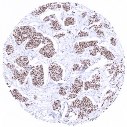
Mouse monoclonal / IgG 1:50 – 1:100 Research Use Only Nuclear Human MSVA-450M GATA3; GATA binding protein-3; GATA-binding factor 3; GATA3; HDR; HDRS; Transacting T-cell-specific transcription factor GATA-3 GATA3 is expressed in breast epithelium, urothelial cells, and a subset of lymphocytes. GATA3 gene at 10p14 consists of 8 exons and codes for GATA3, variant 1, and GATA3, variant 2, proteins of 48 KDa that which differ in one amino acid and may have identical functions. GATA3 is characterized by two conserved zinc finger domains, which bind DNA at the nucleotide sequence GATA. Like other proteins of the GATA family (GATA1-6), GATA3 is a transcription factor that regulates various cellular programs during embryogenesis and differentiation in several organ systems, including parathyroid gland, kidney, breast epithelium, urothelium, and the nervous system. A key role has been identified in T cell development, where GATA3 is the downstream of Notch in differentiation of T-helper cells and important for allergic and humoral immune responses. A role of GATA3 has also been demonstrated for tumor development and progression. GATA3 is frequently found mutated in breast cancer, and downregulation of the protein has been linked to poor prognosis of the disease. GATA3 staining pattern in Normal Tissues with antibody MSVA-450M (images are shown in our “Normal Tissue Gallery”) Brain Cerebrum Negative. Cerebellum Negative. Endocrine Tissues Thyroid Negative. Parathyroid Strong GATA3 positivity in all epithelial cells. Adrenal gland Negative. Pituitary gland Negative. Respiratory system Respiratory epithelium A weak cytoplasmic GATA3 staining can occur in goblet cells of respiratory epithelium (probably non-specific staining). Lung Negative. Gastrointestinal Tract Salivary glands Weak GATA3 staining of glandular cells (mostly mucinous). Esophagus Negative. Stomach A weak cytoplasmic GATA3 staining can occur in gastric glands (probably non-specific staining). Duodenum Negative. Small intestine Negative. Appendix Negative. Colon Negative. Rectum Negative. Liver Negative. Gallbladder Negative. Pancreas Negative. Genitourinary Kidney Strong GATA3 positivity collecting ducts, moderate staining in glomerular podocytes. Urothelium Strong GATA3 positivity... Detectable expression of GATA3 is primarily seen in breast cancer and in urothelial carcinomas. Other tumors for which GATA3 expression has been reported include renal carcinoma, lung cancer and testicular tumors. At low frequency, GATA3 expression can occur in additional tumor entities. The TCGA findings on GATA3 RNA expression in different tumor categories have been summarized in the Human Protein Atlas. Breast cancer of no special type (NST) showing moderate to strong GATA3 immunostaining. (GATA3 immunohistochemistry) Muscle-invasive urothelial carcinoma of the urinary bladder showing a moderate to strong GATA3 positivity of tumor cells. GATA3 negative small cell carcinoma of the lung. Cancer tissue gallery GATA3 ( MSVA-450M) publication summary Relevant publication: Reiswich et al. GATA3 expression in human tumors: A tissue microarray study on 16’557 tumors. Published in Pathobiology 2023 Jan 17:1-14. Epub ahead of print. PMID: 36649695. A total of 13’093 tumors were analyzed from 131 different tumor categories by using the following protocol: Heat-induced antigen retrieval for 5 minutes in an autoclave at 121°C in pH 7,8 Target Retrieval Solution buffer. MSVA-450M at a dilution of 1:50 at 37°C for 60 minutes. Visualization of bound antibody by the EnVision Kit (Dako, Agilent). This protocol was also used for all stainings depicted in our tumor and normal tissue galleries. Overall, 70 (53%) of 131 tumor categories showed detectable GATA3 expression in at least one case and 24 (18%) tumor categories included at least one case with strong GATA3 positivity. The highest positivity rates and the highest levels of expression was found in various categories of breast and urinary bladder neopla... IHC users have different preferences on how the stains should look like. Some prefer high staining intensity of the target stain and even accept some background. Others favor absolute specificity and lighter target stains. Factors that invariably lead to more intense staining include higher concentration of the antibody and visualization tools, longer incubation time, higher temperature during incubation, higher temperature and longer duration of the heat induced epitope retrieval (slide pretreatment). The impact of the pH during slide pretreatment has variable effects and depends on the antibody and the target protein. All images and data shown here and in our image galleries are obtained by the manual protocol described below. Other protocols resulting in equivalent staining are described as well. Manual protocol Freshly cut sections should be used (less than 10 days between cutting and staining). Heat-induced antigen retrieval for 5 minutes in an autoclave at 121°C in pH 7,8 Target ... Because of partly controversial data, the diagnostic utility of GATA3 IHC should be investigated in a large cohort of tumors from different entities. The prognostic role of GATA3 expression in tumor types that are positive in only a fraction of cases is unclear. GATA3 is one of the three genes mutated in >10% of breast cancers (Cancer Genome Atlas) and plays a role in estrogen and androgen receptor signaling. Further studies are needed to elucidate the role, if any, of GATA3 in the development of breast cancer. GATA3 delineates important subgroups of inflammatory cells of which the exact biological and clinical role is not fully understood. GATA3 is thus an important component of antibody panels for use in multicolor immunofluorescence analyses.