Ki-67 (MSVA-267M)
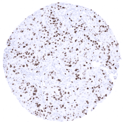
mouse monoclonal / IgG1, kappa 1:100 – 1:200 Research Use Only Nuclear Human MSVA-267M KI-67; Ki67; KI-67 Antigen (KIA); MKI67; Proliferation related Ki-67 antigen tonsil (moderate to strong nuclear staining in 80-90% of the germinal centre B-cells and in the vast majority of the suprabasal squamous epithelial cells). liver (nuclear staining reaction only in scattered hepatocytes (<1%), cytoplasmic staining is absent or only weak) The Ki67 protein is a labile, non-histone nuclear protein expressed in G1, S, G2 and M phases of the cell cycle, then rapidly catabolized at the end of M phase and not detectable in G0 and early G1 cells. When cells enter mitosis, chromosomes undergo chromosome condensation and are coated by a proteinaceous sheath termed the perichromosomal layer (PCL). The PCL comprises approximately one-third of the protein mass of mitotic chromosomes. Ki67 is one of the earliest proteins associated with the PCL and critical for its formation. Ki67 remains on the PCL until telophase.The percentage of Ki67 positive (tumor) cells – also called Ki-67 labeling index (Ki67 LI) – is widely used for quantification of tumor cell proliferation. The Ki67 protein is a labile, non-histone nuclear protein expressed in G1, S, G2 and M phases of the cell cycle, then rapidly catabolized at the end of M phase and not detectable in G0 and early G1 cells. When cells enter mitosis, chromosomes undergo chromosome condensation and are coated by a proteinaceous sheath termed the perichromosomal layer (PCL). The PCL comprises approximately one-third of the protein mass of mitotic chromosomes. Ki67 is one of the earliest proteins associated with the PCL and critical for its formation. Ki67 remains on the PCL until telophase. The percentage of Ki67 positive (tumor) cells – also called Ki-67 labeling index (Ki67 LI) – is widely used for quantification of tumor cell proliferation. Using MSVA-267M , Ki67 positive cells can be seen in the vast majority of tissues. The number of Ki67 positive cells depends on the proliferative activity, which is particularly high in the thymus, germinal centres of lymphoid tissues, and in crypts of intestinal epithelium. These findings obtained by MSVA-267M are largely similar as the protein data described in the Human Protein Atlas (Tissue expression Ki-67) . Suggested positive tissue control: tonsil (moderate to strong nuclear staining in 80-90% of the germinal centre B-cells and in the vast majority of the suprabasal squamous epithelial cells). Suggested negative tissue control: liver (Nuclear staining reaction only in scattered hepatocytes (<1%), cytoplasmic staining is absent or only weak) Placenta, early: Ki67 positive cells are preferentially seen in the storm cells. Lymph node: The highest proliferative activity occurs in germinal centres. Appendix, mucosa: Normal tissue gallery A variable fraction of Ki67 positive cells is seen in virtually every tumor. The percentage of positive tumors is highly variable both within and between tumor entities. The TCGA findings on Ki-67 RNA expression in different tumor categories have been summarized in the Human Protein Atlas. High Ki67 LI in a small cell carcinoma of the lung Low Ki67 LI in papillary thyroid cancer Small cell carcinoma of the urinary bladder with very high Ki67 LI. All tumor cells are Ki67 positive. Cancer tissue gallery No data available at the moment IHC users have different preferences on how the stains should look like. Some prefer high staining intensity of the target stain and even accept some background. Others favor absolute specificity and lighter target stains. Factors that invariably lead to more intense staining include higher concentration of the antibody and visualization tools, longer incubation time, higher temperature during incubation, higher temperature and longer duration of the heat induced epitope retrieval (slide pretreatment). The impact of the pH during slide pretreatment has variable effects and depends on the antibody and the target protein. All images and data shown here and in in our image galleries are obtained by the manual protocol described below. Other protocols resulting in equivalent staining are described as well. Manual protocol Freshly cut TMA sections are deparaffinized and exposed to heat-induced antigen retrieval for 5 minutes in an autoclave at 121°C in pH 9 Target Retrieval Solution buffer.... Ki67 is often used in studies employing multicolor-immunofluorescence. The prognostic role of Ki67 LI is established in many tumor types. Large-scale studies are still needed to exactly determine the practical clinical utility of KI67 measurement. Methods are needed for precise and automated Ki67 measurement in cancer tissues. The fraction of Ki67 positive lymphocyte subpopulations such as for example Ki67 positive CD8 positive cytotoxic T-lymphocytes is under intensive evaluation. The topographical distribution of Ki67 positive lymphocyte subtypes may be relevant. Specificity of MSVA-267M is documented by strong positive nuclear staining in all mitoses and in a large fraction of cells in tissues known to proliferate rapidly. At the same time, staining is always absent in the cytoplasm and on membranes of any cells. The latter especially applies to tissues notorious for showing cytoplasmic background in IHC such as kidney, colonic mucosa, and epidermis. Normal tissue gallery