MCM2 (MSVA-502R)
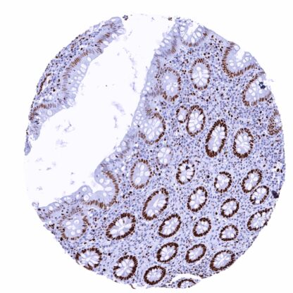
Recombinant Rabbit monoclonal / IgG 1:100 – 1:200 Research Use Only Nucleus Human MSVA-502R DNA replication licensing factor MCM2, Minichromosome maintenance protein 2 homolog, Nuclear protein BM28, BM28; CCNL1; CDC like 1; cdc19; CDCL1; Cell devision cycle like 1; Cyclin like 1; DNA replication licensing factor MCM2; KIAA0030; Minichromosome maintenance complex component 2; Minichromosome maintenance deficient 2 (mitotin); Minichromosome maintenance protein 2 homolog; Mitotin; Nuclear protein BM28 Colon: A strong nuclear MCM2 immunostaining should be seen in virtually all crypt base cells. Colon: MCM2 immunostaining should be less intense or absent in surface epithelial cells and absent in most stroma cells. MCM2 is a highly sensitive marker for proliferating cells. The MCM2 gene is located at 3q21.3 and codes for a nuclear protein which belongs to the highly conserved mini-chromosome maintenance proteins (MCM) 2-7 that play a key role in genome replication. They form a ring-shaped hexameric protein complex which unwinds double-stranded DNA, forms a replication fork during the initiation of DNA replication, and helps to recruit other DNA replication related proteins. The MCM2-7 limits DNA replication to a single occurrence per cell division and is critical for maintaining genome integrity. MCM2 is phosphorylated, and thus regulated by protein kinases such as CDC2 and CDC7. The MCM proteins are expressed in all cells in the G1, S, G2 and M-phase of the cell cycle but in contrast to the better established proliferation marker Ki-67, MCMs are already expressed in early G1 phase. This results in the detection of more proliferating cells as compared to Ki67 immunohistochemistry which might be advantageous in tumor types with low proliferative activity. Images describing the MCM2 staining pattern in normal tissues obtained by the antibody MSVA-502R are shown in our “ Normal Tissue Gallery ”. Brain Cerebrum Negative. Cerebellum Negative. Endocrine Tissues Thyroid Weak to moderate MCM2 staining of a small fraction of follicular cells. Parathyroid MCM2 staining in a small fraction of epithelial cells. Adrenal gland A variable MCM2 staining occurs in a small fraction of adrenocortical cells. Pituitary gland Weak MCM2 staining in a small fraction of cells (neuro- and adenohypophysis). Respiratory system Respiratory epithelium Significant MCM2 staining in a fraction of (mostly basal/suprabasal) respiratory epithelial cells. Lung Strong MCM2 staining of a large subset of pneumocytes. Gastrointestinal Tract Salivary glands Weak MCM2 staining in a fraction of epithelial cells. Esophagus Moderate to strong MCM2 staining of suprabasal and basal cells of the squamous epithelium. Stomach Strong MCM2 immunostaining of many mucous neck cells. Duoden... A nuclear MCM2 immunostaining in a fraction of tumor cells is always seen in cancerous tissues. The TCGA findings on MCM2 RNA expression in different tumor categories have been summarized in the Human Protein Atlas. Adenocarcinoma (Gleason 3+3=6) with weak to moderate MCM2 positivity of fewtumor cells Squamous cell carcinoma with strong MCM2 staining of almost all tumor cells Follicular adenoma with variable MCM2 staining of a fraction of tumor cells Cancer tissue gallery No data available at the moment IHC users have different preferences on how the stains should look like. Some prefer high staining intensity of the target stain and even accept some background. Others favor absolute specificity and lighter target stains. Factors that invariably lead to more intense staining include higher concentration of the antibody and visualization tools, longer incubation time, higher temperature during incubation, higher temperature and longer duration of the heat induced epitope retrieval (slide pretreatment). The impact of the pH during slide pretreatment has variable effects and depends on the antibody and the target protein. All images and data shown here and in our image galleries are obtained by the manual protocol described below. Other protocols resulting in equivalent staining are described as well. Manual protocol Freshly cut sections should be used (less than 10 days between cutting and staining). Heat-induced antigen retrieval for 5 minutes in an autoclave at 121°C in pH 7,8 Target ... The prognostic role of the percentage of MCM2 positive cells is yet unknown. It is unclear whether MCM2 quantification is equally or better suited than the established Ki67-Li for prognosis assessment in tumors with rather low proliferation rate. In principle, there are two ways how the specificity of antibodies can be documented for immunohistochemistry on formalin fixed tissues. These are: 1. Comparison with a second independent method for target expression measurement across a large number of different tissue types (orthogonal strategy), and 2. Comparison with one or several independent antibodies for the same target and showing that all positive staining results are also seen with other antibodies for the same target (independent antibody strategy). For proteins such as MCM2 which are expressed in virtually all tissues but restricted to specific cell types and cell compartments, orthogonal validation is not well suited. However, the comparison of MSVA-502R immunostaining data with data from three independent RNA screening studies, including the Human Protein Atlas (HPA) RNA-seq tissue dataset, the FANTOM5 project, and the Genotype-Tissue Expression (GTEx) project, which are all summarized in the Human Protein Atlas (Tissue ...