MCM7 (MSVA-507R)
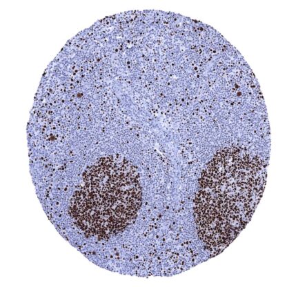
Recombinant Rabbit monoclonal / IgG 1:100 – 1:200 Research Use Only Nuclear Human MSVA-507R CDC47; DNA replication licensing factor MCM7; MCM7 mini chromosome maintenance deficient 7; Minichromosome Maintenance 7; Mini chromosome maintenance protein 7; P1.1-MCM3; P1CDC47; P85MCM; PNAS146 Colon: A strong nuclear MCM7 immunostaining should be seen in virtually all crypt base cells. Colon: MCM7 immunostaining should be largely absent in surface epithelial cells and in most stroma cells. Highly sensitive marker for proliferating cells. The MCM7 gene is located at 7q22.1 and codes for a 80kDa protein which belongs to the highly conserved mini-chromosome maintenance proteins (MCM) 2-7 that play a key role in genome replication. They form a ring-shaped hexameric protein complex which is essential for the pre-replication complex and may be involved in the formation of replication forks, the recruitment of other DNA replication related proteins, and in maintaining genome integrity. Although uncomplexed MCM7 does not have ATPase or DNA helicase activity, it plays a role for DNA helicase activity of the MCM complex. MCM7 expression is regulated by E2F transcription factors under growth factor stimulation by PI3K/AKT, GSK3B , CCND1, and RB1. The MCM proteins are expressed in all cells in the G1, S, G2 and M-phase of the cell cycle but in contrast to the better established proliferation marker Ki-67, MCMs are already expressed in early G1 phase. This results in the detection of more proliferating cells as compared to Ki67 imm... A nuclear MCM7 immunostaining of variable intensity – mostly strong – occurs in the cell compartments of tissues known to contain proliferating cells. This includes suprabasal and (weaker) basal cell layers of squamous epithelium and urothelium, mucous neck cells of the stomach, crypts of the intestine, as well as a fraction of epithelial cells of the gallbladder, respiratory epithelium and the fallopian tube. In the endometrium, almost all epithelial cells and many stromal cells are MCM7 positive but the rate of positive cells decreases in the secretion phase. A particularly strong MCM7 positivity occurs in most cells of germinal centres and the thymic cortex, and also in scattered individual cells all over the lymphatic tissues, while a large fraction of lymphocytes is at least weakly MCM7 positive. Almost all cells of the bone marrow are positive. In the testis, most spermatocytes are positive but mature sperms and probably also spermatogonia are negative. In the placenta, many cell... A nuclear MCM7 immunostaining in a fraction of tumor cells is always seen in cancerous tissues. The TCGA findings on MCM7 RNA expression in different tumor categories have been summarized in the Human Protein Atlas. Penis- Squamous cell carcinoma with strong MCM7 immunostaining of tumor cells Salivary gland- Warthin tumor displaying strong MCM7 staining in few epithelial cells and few lymphocytes Urinary bladder- Muscle-invasive urothelial carcinoma depicting strong MCM7 positivity in all tumor cells Cancer tissue gallery No data available at the moment IHC users have different preferences on how the stains should look like. Some prefer high staining intensity of the target stain and even accept some background. Others favor absolute specificity and lighter target stains. Factors that invariably lead to more intense staining include higher concentration of the antibody and visualization tools, longer incubation time, higher temperature during incubation, higher temperature and longer duration of the heat induced epitope retrieval (slide pretreatment). The impact of the pH during slide pretreatment has variable effects and depends on the antibody and the target protein. All images and data shown here and in our image galleries are obtained by the manual protocol described below. Other protocols resulting in equivalent staining are described as well. Manual protocol Freshly cut sections should be used (less than 10 days between cutting and staining). Heat-induced antigen retrieval for 5 minutes in an autoclave at 121°C in pH 7,8 Target ... Marker for proliferative cells. The prognostic role of the percentage of MCM7 positive cells is yet unknown for most tumor entities. It is unclear whether MCM7 quantification is equally or better suited than the established Ki67-LI for prognosis assessment. In principle, there are two ways how the specificity of antibodies can be documented for immunohistochemistry on formalin fixed tissues. These are: 1. Comparison with a second independent method for target expression measurement across a large number of different tissue types ( orthogonal strategy ), and 2. Comparison with one or several independent antibodies for the same target and showing that all positive staining results are also seen with other antibodies for the same target ( independent antibody strategy ). For proteins such as MCM7 which are expressed in virtually all tissues but restricted to specific cell types and cell compartments, orthogonal validation is not well suited. However, the comparison of MSVA-507R immunostaining data with data from three independent RNA screening studies, including the Human Protein Atlas (HPA) RNA-seq tissue dataset, the FANTOM5 project, and the Genotype-Tissue Expression (GTEx) project, which are all summarized in the Human Protein Atlas (Tis...