Melan A Specific (MSVA-900M)
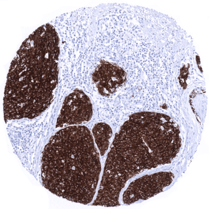
Recombinant Mouse monoclonal / IgG 1:50 – 1:100 Research Use Only Cytoplasmic Human MSVA-900M Antigen LB39-AA, Antigen SK29-AA, Melanoma antigen recognized by T-cells 1, MLAN-A, MLANA Skin: Virtually all melanocytes should show a strong Melan A- immunostaining including a weak to moderate staining in melanocytic dendrites. Kidney: Melan A- staining must be absent in all cells. Melan A Specific is a marker for melanocytes and malignant melanoma. The Melan A (melanocyte antigen) protein, also termed “melanoma antigen recognized by T cells 1” (MART-1) is coded by a gene on chromosome 9p24.1. The 18 kDa protein has a single transmembrane domain and consists of 118 amino acids. The function of the protein is unknown. A small fragment of the protein (about nine amino acids) is bound by MHC class I complexes and presented to cytotoxic T cells. The MART-1/melan A antigen is specific for the melanocyte lineage, found in normal skin, the retina, and melanocytes, but not in other normal tissues. Melan A is thus a highly useful marker for benign and malignant melanocytic tumors. It is of note, that MSVA-900M is fully specific for Melan A. Several other Melan-A antibodies, not only recognize the Melan A protein but – as a result of cross-reactivity – also an unknown structure which is possibly related to corticosteroids. The MSVA antibody MSVA-901M+ (Melan A+) has such properties and can therefore be used as a marker for benign and malign... Using the antibody MSVA-900M (Melan A Specific), a strong staining can be observed in melanocytes in skin and non-keratinizing squamous epithelia from various sites. Staining is absent in all other normal tissues, including the adrenal gland. These findings are largely comparable to the RNA data summarized in the Human Protein Atlas (Tissue expression Melan A) . It is of note, that the different antibodies developed within the protein atlas project contain antibodies of the Melan A+ type showing also strong immunostaining of adrenocortical cells as a result of an antibody . Suggested positive tissue control : Skin: Virtually all melanocytes should show a strong Melan A immunostaining including a weak to moderate staining in melanocytic dendrites. Suggested negative tissue control: Kidney: Melan A staining must be absent in all cells. Skin- A strong Melan-A immunostaining is seen in melanocytes in skin. Melan A staining is absent in the kidney. Using the antibody MSVA-900M (Melan A), i... Melan A is expressed in the vast majority of primary malignant melanomas. Melan A is also expressed in all types of cutaneous naevi and in other tumors of melanocytic differentiation, such as clear cell sarcoma, melanotic neurofibroma, melanotic schwannoma as well as PEComas (perivascular epitheloid cell tumor) including angiomyolipoma, lymphangioleiomyoma(-tosis), and pulmonary sugar tumor. Melan A expression is sometimes reduced and/or only patchy in desmoplastic melanoma and in metastatic melanomas. The TCGA findings on Melan A RNA expression in different tumor categories have been summarized in the Human Protein Atlas. Malignant melanoma of the skin with strong Melan A positivity in all tumor cells Cutaneous malignant melanoma showing diffuse strong Melan A positivity Melan A- negative serous high-grade carcinoma of the ovary. Cancer tissue gallery No data available at the moment IHC users have different preferences on how the stains should look like. Some prefer high staining intensity of the target stain and even accept some background. Others favor absolute specificity and lighter target stains. Factors that invariably lead to more intense staining include higher concentration of the antibody and visualization tools, longer incubation time, higher temperature during incubation, higher temperature and longer duration of the heat induced epitope retrieval (slide pretreatment). The impact of the pH during slide pretreatment has variable effects and depends on the antibody and the target protein. Accordingly, multiple different protocols can generate identical staining results. All images and data shown here and in our image gallery are obtained by the manual protocol described below. Other protocols resulting in equivalent staining are described as well. Manual protocol Freshly cut sections should be used (less than 10 days between cutting and staining). Heat-i... The exact function of Melan A is unknown. The utility of Melan A as a cancer vaccine target is under investigation. Utility of MSVA-900M Melan A is documented by strong positive staining in cell types that are well documented to react with Melan A such as melanocytes and absence of staining in all tissues known to not express Melan A such as adrenal cortex as well as tissues notorious for non-specific IHC background such as kidney, colonic mucosa, and epidermis. Normal tissue gallery