Mesothelin (MSVA-235M)
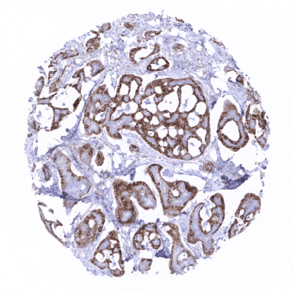
Mouse monoclonal / IgG2 1:100 – 1:200 Research Use Only Cell Surface and Secreted Human MSVA-235M CAK1; Megakaryocyte potentiating factor; Mesothelin; MSLN; SMR; SMRP Tonsil: A fraction of squamous epithelial cells of the tonsil crypts should show strong mesothelin immunostaining. Fallopian tube: An at least moderate mesothelin positivity should be seen at the apical membrane of epithelial cells. Tonsil: Squamous epithelial cells of the tonsil surface and all lymphocytes must not show mesothelin immunostaining. Mesothelin is expressed in various epithelial and mesothelial tumor types. Mesothelin (MSLN) is membranous glycoprotein of 40kDa that is processed by proteolytic cleavage of a larger preprotein encoded by the mesothelin at human chromosome 16p13. MSLN may function as a cell adhesion protein as it is it anchored to the cell membrane and can bind to known cell adhesion molecules such as CA125. However, its exact function has not been clarified as to yet. Mesothelin was originally named after its expression in mesothelial cells. Subsequent work demonstrated that MSLN expression is retained in epithelial mesotheliomas, and that it may also be expressed de-novo in several other cancer types, including ovarian and some kinds of squamous cell carcinomas, making it an attractive target for targeted anti-cancer therapies. [1] [1] Weidemann et al. “Mesothelin Expression in Human Tumors: A Tissue Microarray Study on 12,679 Tumors” Biomedicines 2021, 9, 397. Mesothelin staining pattern in Normal Tissues with antibody MSVA-235M (images are shown in our “Normal Tissue Gallery”) Brain Cerebrum Negative. Cerebellum Negative. Endocrine Tissues Thyroid Negative. Parathyroid Negative. Adrenal gland Negative. Pituitary gland Weak to moderate predominantly cytoplasmic mesothelin staining in the cytoplasm of few cells of the adenohypophysis. Respiratory system Respiratory epithelium Weak to moderate mesothelin staining in goblet cells of respiratory epithelium. Lung Negative. Gastrointestinal Tract Salivary glands Weak to moderate mesothelin staining in some scattered glands in the sublingual gland. Esophagus Negative. Stomach Weak to moderate mesothelin staining in some intermediate (neck) cells of the stomach antrum. Colon Negative. Duodenum Weak to moderate mesothelin staining in some scattered glands in Brunner glands. Rectum Strong mesothelin positivity can be seen of some individual cells and of small cell groups of the rectal mucosa epitheliu... Mesothelin can be found expressed in many different tumor types. The highest rates of positivity (>50% of positive cases) are seen in carcinomas of the ovary, pancreas, endometrium, lung (adenocarcinoma) as well as in malignant mesothelioma. Mesothelin positivity is particularly rare or absent (<5%) carcinomas of the breast, kidney, prostate, and the thyroid as well as in most subtypes of soft tissue tumors. Detailed data on Mesothelin staining by MSVA-235M obtained from an analysis of 12,679 tumors from 122 different tumor types and subtypes have recently been published by Weidemann et at “Mesothelin Expression in Human Tumors: A Tissue Microarray Study on 12,679 Tumors” The TCGA findings on Mesothelin RNA expression in different tumor categories have been summarized in the Human Protein Atlas. Clear cell renal cell carcinoma with moderate to strong cytoplasmic mesothelin positivity. Squamous cell carcinoma of the larynx with strong membranous and cytoplasmic mesothelin staining in th... Mesothelin (MSVA-235M) publication summary Paper used for meta analysis: Weidemann et al “Mesothelin Expression in Human Tumors: A Tissue Microarray Study on 12,679 Tumors” in Biomedicines 2021, 9, 397. A total of 13218 tumors were analyzed from 122 different tumor categories by using the following protocol: Heat-induced antigen retrieval for 5 minutes in an autoclave at 121°C in pH9 Target Retrieval Solution buffer. MSVA-235M at a dilution of 1:150 at 37°C for 60 minutes. Visualization of bound antibody by the EnVision Kit (Dako, Agilent). This protocol was also used for all stainings depicted in our cancer tissue gallery & normal tissue gallery . At least one case with a positive mesothelin immunostaining was seen in 66 (54%) and at least one case with a strong mesothelin immunostaining was seen in 50 (41%) of 122 tumor categories. The distribution of positive staining results is shown in an “organ-systematic” and in a “ranking order” figure below (images based on data from Weideman... IHC users have different preferences on how the stains should look like. Some prefer high staining intensity of the target stain and even accept some background. Others favor absolute specificity and lighter target stains. Factors that invariably lead to more intense staining include higher concentration of the antibody and visualization tools, longer incubation time, higher temperature during incubation, higher temperature and longer duration of the heat induced epitope retrieval (slide pretreatment). The impact of the pH during slide pretreatment has variable effects and depends on the antibody and the target protein. All images and data shown here and in our image galleries are obtained by the manual protocol described below. Other protocols resulting in equivalent staining are described as well. Manual protocol Slides are deparaffinized and exposed to heat-induced antigen retrieval for 5 minutes in an autoclave at 121°C in pH9 buffer. Apply MSVA-235M at a dilution of 1:150 at 37°C ... Mesothelin is a highly promising therapeutic target for which a variety of drugs are under development. A comprehensive study analyzing mesothelin expression in various tumor entities would be helpful to assess the diagnostic significance of mesothelin IHC. Mesothelin expression occurs in a variable fraction of cases in many different tumor types but the prognostic and predictive relevance of mesothelin expression is unclear. The function of mesothelin in cancers is not yet fully elucidated. Specificity of MSVA-235M is documented by strong positive staining in cell types that are well documented to express mesothelin such as tonsil crypts or fallopian tube and absence of staining in all tissues known to not express mesothelin including tissues notorious for non-specific IHC background such as kidney, colonic mucosa, and epidermis. Normal tissue gallery