MRE11 (HMV328)
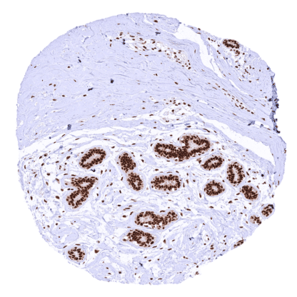
Recombinant Rabbit monoclonal / IgG 1:100 – 1:200 Research Use Only Intracellular Human HMV328 MRE11 homolog, double strand break repair nuclease , ATLD , HNGS1 , MRE11A , MRE11B Colon: A strong nuclear MRE11 staining should be seen in all cells. Cancer with previously documented MRE11 negativity: All cancer cells should remain MRE11 negative while a strong nuclear MRE11 staining should be seen in all non-neoplastic cells. MRE11 is Critical for DNA double strand break repair. Double-strand break repair protein MRE11 (also named MRE11A) is an 80,6 kDa (predominant isoform) protein coded by the MRE11 gene on chromosome 11q21. Together with RAD50 and NBS1, MRE11 forms the MRN complex which is a key element in DNA damage response (DDR) and possesses both single-stranded DNA endonuclease and 3′ to 5′ exonuclease activities. The MRN complex is an early sensor for locating double strand DNA breaks (DSBs) and plays a direct role in both DSB repair and the recruiting of DDR proteins and activation of their downstream signaling. It regulates repair of DNA double-strand breaks in several contexts, including replication, telomere homeostasis, meiosis, apoptosis and immune system development . The MRN complex is involved in multiple different pathways of DSBs repair, including homologous recombination (HR), non-homologous end joining (NHEJ) and the (most error prone) pathway of microhomology-mediated end joining (MMEJ) repair. MRE11 is one of 6 enzymes required for MMEJ... Images describing the MRE11 staining pattern in normal tissues obtained by the antibody HMV328 are shown in our “ Normal Tissue Gallery ”. Brain Cerebrum Distinct nuclear MRE11 staining of all cells. Cerebellum Variable nuclear MRE11 staining of all cells. The staining is strongest in Purkinje cells while it is rather weak in the granule cell layer. Endocrine Tissues Thyroid Strong nuclear MRE11 staining of all cells. Parathyroid Strong nuclear MRE11 staining of all cells. Adrenal gland Distinct nuclear MRE11 staining of all cells. Pituitary gland Distinct nuclear MRE11 staining of all cells. Respiratory system Respiratory epithelium Strong nuclear MRE11 staining of all cells. Lung Strong nuclear MRE11 staining of all cells. Gastrointestinal Tract Salivary glands Strong nuclear MRE11 staining of all cells. Esophagus Strong nuclear MRE11 staining of all cells. Slight decrease of MRE11 staining of squamous epithelium from the basal/suprabasal to the superficial cell layers. Stomach Stron... A variable level of MRE11 staining is seen in the cells of most cancers. Only a few cancers completely lack MRE11 staining. The TCGA findings on MRE11 RNA expression in different tumor categories have been summarized in the Human Protein Atlas. Thyroid – Papillary cancer with strong nuclear MRE11 immunostaining of all tumor cells Ovary – Malignant mixed Mullerian tumor with strong MRE11 positivity of epithelial and mesenchymal tumor cells Colon – Colorectal adenocarcinoma with a marked reduction or loss of MRE11 staining of tumor cells Cancer tissue gallery No data available at the moment IHC users have different preferences on how the stains should look like. Some prefer high staining intensity of the target stain and even accept some background. Others favor absolute specificity and lighter target stains. Factors that invariably lead to more intense staining include higher concentration of the antibody and visualization tools, longer incubation time, higher temperature during incubation, higher temperature and longer duration of the heat induced epitope retrieval (slide pretreatment). The impact of the pH during slide pretreatment has variable effects and depends on the antibody and the target protein. All images and data shown here and in our image galleries are obtained by the manual protocol described below. Other protocols resulting in equivalent staining are described as well. Manual protocol Freshly cut sections should be used (less than 10 days between cutting and staining). Heat-induced antigen retrieval for 5 minutes in an autoclave at 121°C in pH 7,8 Target ... The interplay between MRE11 and its complex network needs to be further evaluated. The predictive role of both decreased and elevated MRE11 expression on cancer radiosensitivity must be further investigated. The clinical potential of the diverse MRE11 inhibitors awaits clarification. There are two ways how the specificity of antibodies can be documented for immunohistochemistry on formalin fixed tissues. These are: 1. Comparison with a second independent method for target expression measurement across a large number of different tissue types (orthogonal strategy), and 2. Comparison with one or several independent antibodies for the same target and showing that all positive staining results are also seen with other antibodies for the same target (independent antibody strategy). Orthogonal validation: For the antibody HMV328 , specificity of its nuclear staining is consistent by the good concordance of the immunostaining data with data from three independent RNA screening studies, including the Human Protein Atlas (HPA) RNA-seq tissue dataset, the FANTOM5 project, and the Genotype-Tissue Expression (GTEx) project, which are all summarized in the Human Protein Atlas (Tissue expression MRE11) . In agreement with HMV328 immunostaining data, MRE11 expression predominated...