MUC2 (HMV310)
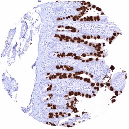
Recombinant Rabbit monoclonal / IgG 1:100 – 1:200 Research Use Only Secreted Human HMV310 mucin 2, oligomeric mucus/gel-forming , MLP , MUC-2 , SMUC Small intestine: A strongMUC2staining should be seen in goblet cells while other epithelial cells as well as inflammatory cells must remain negative. Small intestine:MUC2staining must be absent in all non-goblet cells (non-goblet epithelial cells, stroma cells, inflammatory cells, muscular wall). MUC2 is a main protein of intestinal mucus barrier. Mucin 2 (MUC2) is an oligomeric mucus gel-forming protein coded by the MUC2 gene at chromosome 11p15.5. The protein is secreted by intestinal goblet cells to the gut where it constitutes the main component of colorectal mucus. The mucins function as a defense mechanism to maintain the integrity of the epithelial cells which are continuously exposed to luminal contents that include large quantities of different bacteria, proteases, bile, and ingested toxins. The mucin barrier consists of two layers. The inner layer is directly attached to the epithelium, densely packed, largely consists of uncleaved MUC2, and is free from bacterial colonization. The outer layer contains bacteria and is less dense due of proteolytic cleavage of MUC2. Under normal conditions, the protease-resistant mucus layer prevents intestinal bacteria to make physical contact with epithelial cells by virtue of its only minute pore sizes and strong hydrophobic properties capable to repel bacteria in the aqueous lumen. ... Images describing the MUC2 staining pattern in normal tissues obtained by the antibody HMV310 are shown in our “ Normal Tissue Gallery ”. Brain Cerebrum Negative. Cerebellum Negative. Endocrine Tissues Thyroid Negative. Parathyroid Negative. Adrenal gland Negative. Pituitary gland Negative. Respiratory system Respiratory epithelium Negative. Lung Negative. Gastrointestinal Tract Salivary glands Negative. Esophagus Negative. Stomach Negative. Colon Strong MUC2 positivity of mucins in goblet cells. Duodenum Strong MUC2 positivity of mucins in goblet cells. Rectum Strong MUC2 positivity of mucins in goblet cells. Small intestine Strong MUC2 positivity of mucins in goblet cells. Liver Negative. Gallbladder Negative. Pancreas Negative. Genitourinary Kidney Negative. Urothelium Negative. Male genital Prostate Negative. Seminal vesicles Negative. Testis Negative. Epididymis Negative. Female genital Breast Negative. Uterus, myometrium Negative. Uterus, ectocervix Negative. Uterus endocervix Ne... MUC2 is preferably expressed in a fraction of colorectal adenocarcinomas: It also occurs in breast and stomach cancers as well as (less commonly) in many other tumor entities (preferably adenocarcinoma). The TCGA findings on MUC2 RNA expression in different tumor categories have been summarized in the Human Protein Atlas. Muscle-invasive urothelial carcinoma with intense
MUC2 immunostaining of all tumor cells.strong MUC2 staining of virtually all tumor cells. Kidney: MUC2 negative papillary renal cell carcinoma. Invasive breast cancer of no special type (NST) with
strong MUC2 staining of virtually all tumor cells. Cancer tissue gallery No data available at the moment IHC users have different preferences on how the stains should look like. Some prefer high staining intensity of the target stain and even accept some background. Others favor absolute specificity and lighter target stains. Factors that invariably lead to more intense staining include higher concentration of the antibody and visualization tools, longer incubation time, higher temperature during incubation, higher temperature and longer duration of the heat induced epitope retrieval (slide pretreatment). The impact of the pH during slide pretreatment has variable effects and depends on the antibody and the target protein. All images and data shown here and in our image galleries are obtained by the manual protocol described below. Other protocols resulting in equivalent staining are described as well. Manual protocol Freshly cut sections should be used (less than 10 days between cutting and staining). Heat-induced antigen retrieval for 5 minutes in an autoclave at 121°C in pH 7,8 Target ... The prognostic relevance of MUC2 expression in tumors is not fully clarified. The potential predictive role of MUC2 expression measurement in tumors needs to be further investigated. There are two ways how the specificity of antibodies can be documented for immunohistochemistry on formalin fixed tissues. These are: 1. Comparison with a second independent method for target expression measurement across a large number of different tissue types (orthogonal strategy), and 2. Comparison with one or several independent antibodies for the same target and showing that all positive staining results are also seen with other antibodies for the same target (independent antibody strategy). Orthogonal validation: For the antibody HMV310 specificity is suggested by the complete concordance of the immunostaining data with data from three independent RNA screening studies, including the Human Protein Atlas (HPA) RNA-seq tissue dataset, the FANTOM5 project, and the Genotype-Tissue Expression (GTEx) project, which are all summarized in the human protein atlas Human Protein Atlas (Tissue expression MUC2) . MUC2 positivity by HMV310 is only detectable in the tissues with documented MUC...