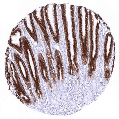
MUC5AC (MSVA-109M)
mouse monoclonal / IgG1 1:100 – 1:200 Research Use Only Cytoplasmic Human MSVA-109M Apomucin Major Airway Glycoprotein, Mucin 5 subtype AC tracheobronchial, Mucin 5 Subtypes A and C, Mucin 5AC oligomeric mucus/gel forming, Tracheobronchial Mucin Gastric mucosa: A strong cytoplasmic MUC5AC staining should be seen in the surface epithelium. Kidney: MUC5AC staining should be absent. Mucin 5AC (MUC5AC) is one of five gel-forming mucins that are found in humans. It is encoded by the mucin cluster at chromosome 11p15, which also encodes the genes of the related mucins (MUC2, -5B, -6, and -19). The large (641 KDa) protein harbors multiple polymerization sites enabling formation of the mucin gel layer. Mucin layers are found at the surface of many epithelia, including those of the upper and lower respiratory tract, stomach, and endocervix, where they the build a barrier against chemical and mechanical damage. Microbial pathogens clotted in the mucin will be removed by the mucociliary system. Recent work suggests that MUC5AC may also play a role for tumor development and progression of lung, colorectal and pancreatic cancers, as it interacts with tumor relevant proteins such as p53, integrin ß4 and ß-catenin. In colorectal cancer, MUC5AC expression is independently linked to both right colon location and microsatellite instability. Mucin 5AC (MUC5AC) is one of five gel-forming mucins that are found in humans. It is encoded by the mucin cluster at chromosome 11p15, which also encodes the genes of the related mucins (MUC2, -5B, -6, and -19). The large (641 KDa) protein harbors multiple polymerization sites enabling formation of the mucin gel layer. Mucin layers are found at the surface of many epithelia, including those of the upper and lower respiratory tract, stomach, and endocervix, where they the build a barrier against chemical and mechanical damage. Microbial pathogens clotted in the mucin will be removed by the mucociliary system. Recent work suggests that MUC5AC may also play a role for tumor development and progression of lung, colorectal and pancreatic cancers, as it interacts with tumor relevant proteins such as p53, integrin ß4 and ß-catenin. In colorectal cancer, MUC5AC expression is independently linked to both right colon location and microsatellite instability. MUC5AC staining pattern in Normal Tissues with antibody MSVA-109M (images are shown in our “Normal Tissue Gallery”) Brain Cerebrum Negative. Cerebellum Negative. Endocrine Tissues Thyroid Negative. Parathyroid Negative. Adrenal gland Negative. Pituitary gland Negative. Respiratory system Respiratory epithelium Strong cytoplasmic staining of goblet cells. Lung Negative. Gastrointestinal Tract Salivary glands Negative. Esophagus Negative. Stomach Strong cytoplasmic MUC5AC staining in all cells of the stomach surface epithelium. Colon Few scattered MUC5AC positive cells do occur. Duodenum Few scattered MUC5AC positive cells do occur. Rectum Few scattered MUC5AC positive cells do occur. Small intestine Few scattered MUC5AC positive cells do occur. Liver Negative. Gallbladder Moderate to strong cytoplasmic MUC5AC staining of the surface epithelium may be focal. Pancreas Negative. Genitourinary Kidney Negative. Urothelium Negative. Male genital Prostate Negative. Seminal vesicles Negative. T... In principle, three different patterns can be seen: diffuse staining of all cells, patchy focal staining of tumor areas that sometimes exhibit a different morphology as compared to non-stained areas groups, and a mosaic pattern containing a variable number of scattered strongly MUC5AC positive cancer cells quite regularly distributed between clearly negative tumor cells. MUC5AC expression is most commonly seen in adenocarcinomas derived from esophagus (>70%), pancreas (>60%), and the stomach (>40%). At a frequency below 30% it is also seen in various other adenocarcinomas such as endometrial cancer, colorectal carcinoma,cholangiocellular carcinoma, and lung adenocarcinoma. At a frequency below 15% MUC5AC positivity is also seen in various other tumor types including squamous cell carcinoma of the lung, neuroendocrine cancer, large cell lung cancer. MUC5AC is rarely or never present in various important other tumor types such as in cancers of the breast, prostate, kidney, serous ovarian... MUC5AC (MSVA-109M) publication summary Papers used for data compilation: Rico SD et al. “MUC5AC Expression in Various Tumor Types and Nonneoplastic Tissue: A Tissue Microarray Study on 10 399 Tissue Samples.” Published in Technol Cancer Res Treat. Jan-Dec 2021 PMID: 34547930 Rico SD et al. “MUC5AC expression is linked to mucinous/endometroid subtype, absence of nodal metastasis and mismatch repair deficiency in ovarian cancer.” Published in Pathol Res Pract. 2021 Aug; 224:153533 PMID: 34171599 Rico SD et al. “Elevated MUC5AC expression is associated with mismatch repair deficiency and proximal tumor location but not with cancer progression in colon cancer.” Published in Med Mol Morphol. 2021 Jun;54(2): 156-165 PMID: 33373033 In these three studies, a total of 10210 tumors were analyzed from 114 different tumor categories by using the following protocol: Heat-induced antigen retrieval for 5 minutes in an autoclave at 121°C in pH 7.8 Target Retrieveal Solution buffer. MSVA-109M at a dil... IHC users have different preferences on how the stains should look like. Some prefer high staining intensity of the target stain and even accept some background. Others favor absolute specificity and lighter target stains. Factors that invariably lead to more intense staining include higher concentration of the antibody and visualization tools, longer incubation time, higher temperature during incubation, higher temperature and longer duration of the heat induced epitope retrieval (slide pretreatment). The impact of the pH during slide pretreatment has variable effects and depends on the antibody and the target protein. Accordingly, multiple different protocols can generate identical staining results. All images and data shown here are and in our image galleries were obtained by the manual protocol described below. Other protocols resulting in equivalent staining are described as well. Manual protocol Freshly cut sections should be used (less than 10 days between cutting and staining).... A comprehensive study analyzing MUC5AC expression in various different tumor entities would be helpful to assess the diagnostic significance of MUC5AC IHC. The prognostic relevance of MUC5AC expression and of different immunostaining patterns (diffuse vs “mosaic”) should be evaluated. The use of serum MUC5AC measurement as a putative instrument for monitoring response to therapy and early detection of recurrence in case of MUC5AC positive primary tumors should be investigated. Specificity of MSVA-109M is documented by strong positive staining in cell types that are well documented to express MUC5AC such as surface epithelium of the stomach and bronchial epithelium goblet cells and absence of staining in all tissues known to not express MUC5AC including tissues notorious for non-specific IHC background such as kidney, colonic mucosa, and epidermis. Normal tissue gallery