MUC6 (MSVA-806R)
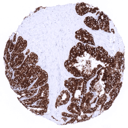
Recombinant Rabbit monoclonal / IgG1 1:100 – 1:200 Research Use Only Cytoplasmic Human MSVA-806R Gastric mucin 6; MUC6; MUC6 mucin; Mucin 6 oligomeric mucus/gel forming; Mucin glycoprotein Fragment; Mucin-6; Secretory mucin MUC6 Stomach: Mucous secreting glands should be strongly positive. Stomach: Surface epithelium should be completely negative. Mucin 6 (MUC6) is a secretory gastric mucin which is expressed in various tumor types. Mucin 6 ( MUC6) is a secretory gastric mucin that was first identified from a gastric mucosal cDNA library. As a secretory mucin, its main role is the formation of a protective barrier for the stomach epithelium and the lubrification of nutrition which is accomplished in collaboration with other mucins. MUC6 is also expressed in various cancers, where it may also play a role for tumor biology. There is evidence that MUC6 inhibits tumor cell motility and that it becomes epigenetically deregulated in carcinoma cells. The strongest MUC6 expression (+++) is detected in epithelial cells of seminal vesicles, Brunner glands of the duodenum, and the mucous secreting glands – but not the surface epithelium – of the stomach. MUC6 immunostaining is also consistently present in Intercalated and interlobular ducts of the pancreas, small juxtaportal bile ducts (portal bile ducts are largely negative), gallbladder surface epithelium (not all cells in all samples), epididymis (more intense staining in the cauda than in the caput, where positivity can be focal), fallopian tube (variable number of scattered positive cells), and endocervical glands (weak). In some samples, few scattered MUC6 positive cells can be found in collecting ducts of the kidney, breast glands, pregnancy endometrium, and in the trophoblast of the first trimenon placenta. Skin and non-keratinizing squamous epithelium of oral cavity, lip, tonsil surface and crypts, ectocervix, and esophagus as well as Hassal’s corpuscles in the thymus are MUC6... MUC6 can be expressed in various tumor types including for example pancreatic, breast, ovarian, endometrial, stomach, colorectal, and cholangiocellular carcinoma. In principle, three different patterns can be seen: diffuse staining of all cells, patchy focal staining of tumor areas that sometimes exhibit a different morphology as compared to non-stained areas groups, and a mosaic pattern containing a variable number of scattered strongly MUC6 positive cancer cells quite regularly distributed between clearly negative tumor cells. The TCGA findings on MUC6 RNA expression in different tumor categories have been summarized in the Human Protein Atlas. Strong MUC6 staining in all cancer cells of a gastric adenocarcinoma (intestinal type). Strong MUC6 immunostaining in a fraction of cells in a ductal adenocarcinoma of the pancreas. Strong MUC6 immunostaining in an invasive breast carcinoma of no special type (NST). Cancer tissue gallery MUC6 (MSVA-806R) publication summary Relevant publication: Dwertmann Rico et al. “Pattern of MUC6 expression across 119 different tumor types: A tissue microarray study on 15 412 tumors” Published in Pathol Int. 2023 Jul;73(7):281-296. PMID: 37057870 A total of 11,685 tumors from 119 different tumor categories were successfully analyzed by using the following protocol: Heat-induced antigen retrieval for 5 minutes in an autoclave at 121°C in pH 7,8 Target Retrieval Solution buffer. MSVA-806R, at a dilution of 1:150 at 37°C for 60 minutes. Visualization of bound antibody by the EnVision Kit (Dako, Agilent). This protocol was also used for all stainings depicted in our tumor and normal tissue galleries. Overall, 50 of 119 tumor categories showed a detectable MUC6 staining of at least one tumor with 33 tumor categories showing at least in one case a strong positivity. The highest positivity rates occurred in mucinous carcinomas of the breast (44%), gastric adenocarcinomas (30%–40%), esopha... IHC users have different preferences on how the stains should look like. Some prefer high staining intensity of the target stain and even accept some background. Others favor absolute specificity and lighter target stains. Factors that invariably lead to more intense staining include higher concentration of the antibody and visualization tools, longer incubation time, higher temperature during incubation, higher temperature and longer duration of the heat induced epitope retrieval (slide pretreatment). The impact of the pH during slide pretreatment has variable effects and depends on the antibody and the target protein. All images and data shown here and in our image galleries are obtained by the manual protocol described below. Other protocols resulting in equivalent staining are described as well. Manual protocol Freshly cut sections should be used (less than 10 days between cutting and staining). Heat-induced antigen retrieval for 5 minutes in an autoclave at 121°C in pH 7,8 Target ... A comprehensive study analyzing MUC6 expression in various different tumor entities would be helpful to assess the diagnostic significance of MUC6 IHC. The clinical significance of MUC6 expression in various cancer entities is unclear. Specificity of MSVA-806R is documented by strong positive staining in cell types that are well documented to express MUC6 such as glandular but not surface epithelium of the stomach, Intercalated and interlobular ducts of the pancreas, and seminal vesicle. The complete absence of MUC6 immunostaining in cell types from 70 different normal tissue types documents absence of cross-reactivity and low propensity to generate non-specific background staining for MSVA-806R . Normal tissue gallery